 Movie
Movie Controller

[English] 日本語
 Yorodumi
- PDB-8byg: fragment-linked stabilizer for ERa - 14-3-3 interaction (1047648) -
+ Open data

- Basic information
| Entry | Database: PDB / ID: 8byg | |||||||||
|---|---|---|---|---|---|---|---|---|---|---|
| Title | fragment-linked stabilizer for ERa - 14-3-3 interaction (1047648) | |||||||||
 Components Components |
| |||||||||
 Keywords Keywords |  STRUCTURAL PROTEIN / 14-3-3 / ERa / fragment linking / stabilization STRUCTURAL PROTEIN / 14-3-3 / ERa / fragment linking / stabilization | |||||||||
| Function / homology |  Function and homology information Function and homology informationregulation of epidermal cell division / protein kinase C inhibitor activity / positive regulation of epidermal cell differentiation / keratinocyte development / keratinization / Regulation of localization of FOXO transcription factors / keratinocyte proliferation / phosphoserine residue binding / Activation of BAD and translocation to mitochondria / negative regulation of keratinocyte proliferation ...regulation of epidermal cell division / protein kinase C inhibitor activity / positive regulation of epidermal cell differentiation / keratinocyte development / keratinization / Regulation of localization of FOXO transcription factors / keratinocyte proliferation / phosphoserine residue binding / Activation of BAD and translocation to mitochondria / negative regulation of keratinocyte proliferation / establishment of skin barrier / SARS-CoV-2 targets host intracellular signalling and regulatory pathways / Chk1/Chk2(Cds1) mediated inactivation of Cyclin B:Cdk1 complex / protein kinase A signaling / negative regulation of stem cell proliferation / SARS-CoV-1 targets host intracellular signalling and regulatory pathways / RHO GTPases activate PKNs / protein export from nucleus / negative regulation of innate immune response / protein sequestering activity / TP53 Regulates Transcription of Genes Involved in G2 Cell Cycle Arrest / release of cytochrome c from mitochondria / positive regulation of protein export from nucleus / stem cell proliferation / Translocation of SLC2A4 (GLUT4) to the plasma membrane / TP53 Regulates Metabolic Genes / negative regulation of protein kinase activity / negative regulation of cysteine-type endopeptidase activity involved in apoptotic process / intrinsic apoptotic signaling pathway in response to DNA damage / positive regulation of cell growth / regulation of cell cycle / cadherin binding / protein kinase binding / negative regulation of transcription by RNA polymerase II / signal transduction / extracellular space / extracellular exosome / identical protein binding / nucleus / cytosol / cytoplasmSimilarity search - Function | |||||||||
| Biological species |  Homo sapiens (human) Homo sapiens (human) | |||||||||
| Method | X-RAY DIFFRACTION / MOLECULAR REPLACEMENT / Resolution: 1.7 Å | |||||||||
 Authors Authors | Visser, E.J. / Sijbesma, E. / Ottmann, C. | |||||||||
| Funding support |  Netherlands, 1items Netherlands, 1items
| |||||||||
 Citation Citation | Journal: Angew.Chem.Int.Ed.Engl. / Year: 2023 Title: From Tethered to Freestanding Stabilizers of 14-3-3 Protein-Protein Interactions through Fragment Linking. Authors: Visser, E.J. / Jaishankar, P. / Sijbesma, E. / Pennings, M.A.M. / Vandenboorn, E.M.F. / Guillory, X. / Neitz, R.J. / Morrow, J. / Dutta, S. / Renslo, A.R. / Brunsveld, L. / Arkin, M.R. / Ottmann, C. | |||||||||
| History |
|
- Structure visualization
| Structure viewer | Molecule: MolmilJmol/JSmol |
|---|
- Downloads & links
-Download
| PDBx/mmCIF format | 8byg.cif.gz | 112.3 KB | Display | PDBx/mmCIF format |
|---|---|---|---|---|
| PDB format | pdb8byg.ent.gz | 84.8 KB | Display | PDB format |
| PDBx/mmJSON format | 8byg.json.gz | Tree view | PDBx/mmJSON format | |
| Others |  Other downloads Other downloads |
-Validation report
| Arichive directory | https://data.pdbj.org/pub/pdb/validation_reports/by/8bygftp://data.pdbj.org/pub/pdb/validation_reports/by/8byg | HTTPS FTP |
|---|
-Related structure data
| Related structure data | 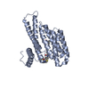 8bwjC 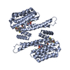 8bwxC 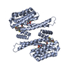 8bwzC  8bx0C 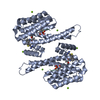 8bx3C 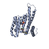 8bx4C 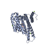 8bxiC  8bxmC 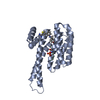 8bxnC 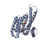 8bxoC 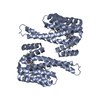 8bxqC 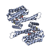 8bxsC  8by9C 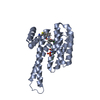 8bybC  8bycC 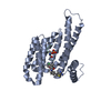 8bydC 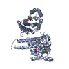 8byeC 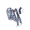 8byfC 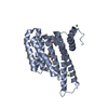 8byoC 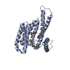 8byyC  8byzC 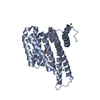 8bz0C 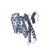 8bz9C 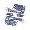 8bzaC  8bzbC  8bzwC  8c04C  8c0kC 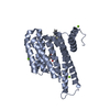 8c4fC 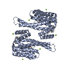 8c4gC C: citing same article ( |
|---|---|
| Similar structure data |
-Links
 PDBj
PDBj


- Assembly
| Deposited unit | 
| ||||||||
|---|---|---|---|---|---|---|---|---|---|
| 1 | 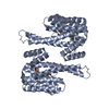
| ||||||||
| Unit cell |
|
-Components
| #1: Protein | Mass: 26542.914 Da / Num. of mol.: 1 Source method: isolated from a genetically manipulated source Source: (gene. exp.) Homo sapiens (human) / Gene: SFN, HME1 / Production host:  Escherichia coli (E. coli) / References: UniProt: P31947 Escherichia coli (E. coli) / References: UniProt: P31947 |
|---|---|
| #2: Protein/peptide | Mass: 613.596 Da / Num. of mol.: 1 / Source method: obtained synthetically / Source: (synth.) Homo sapiens (human) |
| #3: Chemical | ChemComp-S6O / ~{  Mass: 459.004 Da / Num. of mol.: 1 / Source method: obtained synthetically / Formula: C23H27ClN4O2S Mass: 459.004 Da / Num. of mol.: 1 / Source method: obtained synthetically / Formula: C23H27ClN4O2S |
| #4: Water | ChemComp-HOH / Water Mass: 18.015 Da / Num. of mol.: 335 / Source method: isolated from a natural source / Formula: H2O Mass: 18.015 Da / Num. of mol.: 335 / Source method: isolated from a natural source / Formula: H2O |
| Has ligand of interest | Y |
-Experimental details
-Experiment
| Experiment | Method: X-RAY DIFFRACTION / Number of used crystals: 1 |
|---|
- Sample preparation
| Crystal | Density Matthews: 2.67 Å3/Da / Density % sol: 53.85 % |
|---|---|
| Crystal grow | Temperature: 277 K / Method: vapor diffusion, sitting drop Details: 0.095 M HEPES (pH 7.1), PEG400 (24% (v/v)), 0.19 M CaCl2 and 5% (v/v) Glycerol |
-Data collection
| Diffraction | Mean temperature: 100 K / Serial crystal experiment: N |
|---|---|
| Diffraction source | Source: SEALED TUBE / Type: RIGAKU MICROMAX-003 / Wavelength: 1.54187 Å |
| Detector | Type: DECTRIS PILATUS 200K / Detector: PIXEL / Date: Jul 26, 2019 |
| Radiation | Protocol: SINGLE WAVELENGTH / Monochromatic (M) / Laue (L): M / Scattering type: x-ray |
| Radiation wavelength | Wavelength: 1.54187 Å / Relative weight: 1 |
| Reflection | Resolution: 1.7→66.36 Å / Num. obs: 189444 / % possible obs: 96.6 % / Redundancy: 6.1 % / CC1/2: 0.996 / Net I/σ(I): 11.6 |
| Reflection shell | Resolution: 1.7→1.73 Å / Num. unique obs: 1263 / CC1/2: 0.488 |
- Processing
| Software |
| |||||||||||||||||||||||||||||||||||||||||||||||||||||||||||||||||||||||||||||||||||||||||||
|---|---|---|---|---|---|---|---|---|---|---|---|---|---|---|---|---|---|---|---|---|---|---|---|---|---|---|---|---|---|---|---|---|---|---|---|---|---|---|---|---|---|---|---|---|---|---|---|---|---|---|---|---|---|---|---|---|---|---|---|---|---|---|---|---|---|---|---|---|---|---|---|---|---|---|---|---|---|---|---|---|---|---|---|---|---|---|---|---|---|---|---|---|
| Refinement | Method to determine structure: MOLECULAR REPLACEMENT / Resolution: 1.7→34.12 Å / SU ML: 0.2 / Cross valid method: FREE R-VALUE / σ(F): 1.34 / Phase error: 22.24 / Stereochemistry target values: ML
| |||||||||||||||||||||||||||||||||||||||||||||||||||||||||||||||||||||||||||||||||||||||||||
| Solvent computation | Shrinkage radii: 0.9 Å / VDW probe radii: 1.11 Å / Solvent model: FLAT BULK SOLVENT MODEL | |||||||||||||||||||||||||||||||||||||||||||||||||||||||||||||||||||||||||||||||||||||||||||
| Refinement step | Cycle: LAST / Resolution: 1.7→34.12 Å
| |||||||||||||||||||||||||||||||||||||||||||||||||||||||||||||||||||||||||||||||||||||||||||
| Refine LS restraints |
| |||||||||||||||||||||||||||||||||||||||||||||||||||||||||||||||||||||||||||||||||||||||||||
| LS refinement shell |
|
