[English] 日本語
Yorodumi
- PDB-8bto: Helical structure of BcThsA in complex with 1''-3'gcADPR -

+
Open data


ID or keywords:

Loading...

-
Basic information

Entry
Database: PDB / ID: 8bto
TitleHelical structure of BcThsA in complex with 1''-3'gcADPR
ComponentsNAD(+) hydrolase ThsA
KeywordsHYDROLASE / Thoeris / SIR2 domain / SLOG domain / 3'cADPR
Function / homology
Function and homology information


NAD+ glycohydrolase / defense response to virus / hydrolase activity / nucleotide binding / cytoplasm
Similarity search - Function
NAD(+) hydrolase ThsA, Sir2/TIR-associating SLOG domain / Sir2- and TIR-associating SLOG family / SIR2-like domain / SIR2-like domain / Sirtuin family, catalytic core domain / Sirtuin catalytic domain profile. / DHS-like NAD/FAD-binding domain superfamily
Similarity search - Domain/homology
NICOTINAMIDE-ADENINE-DINUCLEOTIDE / Chem-OJC / NAD(+) hydrolase ThsA
Similarity search - Component
Biological speciesBacillus cereus MSX-D12 (bacteria)
MethodELECTRON MICROSCOPY / helical reconstruction / cryo EM / Resolution: 2.96 Å
AuthorsTamulaitiene, G. / Sasnauskas, G. / Sabonis, D.
Funding supportLithuania, 1items
OrganizationGrant numberCountry
Research Council of LithuaniaS-MIP-21-6Lithuania
Citation
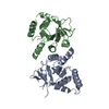
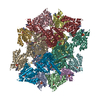
Journal: Nature / Year: 2024
Title: Activation of Thoeris antiviral system via SIR2 effector filament assembly.
Authors: Giedre Tamulaitiene / Dziugas Sabonis / Giedrius Sasnauskas / Audrone Ruksenaite / Arunas Silanskas / Carmel Avraham / Gal Ofir / Rotem Sorek / Mindaugas Zaremba / Virginijus Siksnys /
Abstract: To survive bacteriophage (phage) infections, bacteria developed numerous anti-phage defence systems. Some of them (for example, type III CRISPR-Cas, CBASS, Pycsar and Thoeris) consist of two modules: ...To survive bacteriophage (phage) infections, bacteria developed numerous anti-phage defence systems. Some of them (for example, type III CRISPR-Cas, CBASS, Pycsar and Thoeris) consist of two modules: a sensor responsible for infection recognition and an effector that stops viral replication by destroying key cellular components. In the Thoeris system, a Toll/interleukin-1 receptor (TIR)-domain protein, ThsB, acts as a sensor that synthesizes an isomer of cyclic ADP ribose, 1''-3' glycocyclic ADP ribose (gcADPR), which is bound in the Smf/DprA-LOG (SLOG) domain of the ThsA effector and activates the silent information regulator 2 (SIR2)-domain-mediated hydrolysis of a key cell metabolite, NAD (refs. ). Although the structure of ThsA has been solved, the ThsA activation mechanism remained incompletely understood. Here we show that 1''-3' gcADPR, synthesized in vitro by the dimeric ThsB' protein, binds to the ThsA SLOG domain, thereby activating ThsA by triggering helical filament assembly of ThsA tetramers. The cryogenic electron microscopy (cryo-EM) structure of activated ThsA revealed that filament assembly stabilizes the active conformation of the ThsA SIR2 domain, enabling rapid NAD depletion. Furthermore, we demonstrate that filament formation enables a switch-like response of ThsA to the 1''-3' gcADPR signal.
History
DepositionNov 29, 2022Deposition site: PDBE / Processing site: PDBE
Revision 1.0Feb 21, 2024Provider: repository / Type: Initial release
Revision 1.1Mar 6, 2024Group: Database references / Category: citation / citation_author / Item: _citation.pdbx_database_id_PubMed / _citation.title
Revision 1.2Mar 27, 2024Group: Database references / Category: citation
Item: _citation.journal_volume / _citation.page_first / _citation.page_last
Revision 1.3Apr 3, 2024Group: Database references / Category: citation_author / Item: _citation_author.identifier_ORCID

-
Structure visualization

Structure viewerMolecule:
MolmilJmol/JSmol

Downloads & links

-
Assembly

Deposited unit
A: NAD(+) hydrolase ThsA
B: NAD(+) hydrolase ThsA
C: NAD(+) hydrolase ThsA
D: NAD(+) hydrolase ThsA
E: NAD(+) hydrolase ThsA
F: NAD(+) hydrolase ThsA
G: NAD(+) hydrolase ThsA
H: NAD(+) hydrolase ThsA
I: NAD(+) hydrolase ThsA
J: NAD(+) hydrolase ThsA
K: NAD(+) hydrolase ThsA
L: NAD(+) hydrolase ThsA
hetero molecules


Theoretical massNumber of molelcules
Total (without water)679,47036
Polymers665,01312
Non-polymers14,45724
Water0
1


  • Idetical with deposited unit
  • defined by author
  • Evidence: light scattering, DLS
TypeNameSymmetry operationNumber
identity operation1_5551
Buried area70000 Å2
ΔGint-76 kcal/mol
Surface area209260 Å2

-
Components

#1: Protein
NAD(+) hydrolase ThsA / NADase ThsA / Thoeris protein ThsA


Mass: 55417.746 Da / Num. of mol.: 12 / Mutation: N112A
Source method: isolated from a genetically manipulated source
Source: (gene. exp.) Bacillus cereus MSX-D12 (bacteria) / Gene: thsA, II9_05448 / Production host: Escherichia coli BL21(DE3) (bacteria) / References: UniProt: J8G6Z1, NAD+ glycohydrolase
#2: Chemical
ChemComp-OJC / (2R,3R,3aS,5S,6R,7S,8R,11R,13S,15aR)-2-(6-amino-9H-purin-9-yl)-3,6,7,11,13-pentahydroxyoctahydro-2H,5H,11H,13H-5,8-epoxy-11lambda~5~,13lambda~5~-furo[2,3-g][1,3,5,9,2,4]tetraoxadiphosphacyclotetradecine-11,13-dione


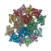
Mass: 541.300 Da / Num. of mol.: 12 / Source method: obtained synthetically / Formula: C15H21N5O13P2 / Feature type: SUBJECT OF INVESTIGATION
#3: Chemical
ChemComp-NAD / NICOTINAMIDE-ADENINE-DINUCLEOTIDE / Nicotinamide adenine dinucleotide


Mass: 663.425 Da / Num. of mol.: 12 / Source method: obtained synthetically / Formula: C21H27N7O14P2 / Feature type: SUBJECT OF INVESTIGATION / Comment: NAD*YM
Has ligand of interestY

-
Experimental details

-
Experiment

ExperimentMethod: ELECTRON MICROSCOPY
EM experimentAggregation state: HELICAL ARRAY / 3D reconstruction method: helical reconstruction

-
Sample preparation

ComponentName: BcThsA in complex with 1''-3'gcADPR / Type: COMPLEX / Entity ID: #1 / Source: RECOMBINANT
Molecular weightExperimental value: NO
Source (natural)Organism: Bacillus cereus (bacteria)
Source (recombinant)Organism: Escherichia coli (E. coli)
Buffer solutionpH: 7.5
Buffer component
IDConc.NameBuffer-ID
120 mMNa-Hepes1
2150 mMsodium chloride1
31 mMDTT1
SpecimenConc.: 1 mg/ml / Embedding applied: NO / Shadowing applied: NO / Staining applied: NO / Vitrification applied: YES
Specimen supportGrid material: COPPER / Grid mesh size: 300 divisions/in. / Grid type: Quantifoil R1.2/1.3
VitrificationInstrument: FEI VITROBOT MARK IV / Cryogen name: ETHANE / Humidity: 95 % / Chamber temperature: 277 K

-
Electron microscopy imaging

MicroscopyModel: TFS GLACIOS
Electron gunElectron source: FIELD EMISSION GUN / Accelerating voltage: 200 kV / Illumination mode: OTHER
Electron lensMode: BRIGHT FIELDBright-field microscopy / Nominal defocus max: 2000 nm / Nominal defocus min: 1000 nm
Specimen holderCryogen: NITROGEN
Image recordingAverage exposure time: 46.33 sec. / Electron dose: 30.64 e/Å2 / Detector mode: COUNTING / Film or detector model: FEI FALCON III (4k x 4k) / Num. of grids imaged: 1 / Num. of real images: 2321

-
Processing

Software
NameVersionClassification
phenix.real_space_refine1.19.2_4158refinement
PHENIX1.19.2_4158refinement
EM software
IDNameCategory
2EPUimage acquisition
7UCSF ChimeraXmodel fitting
9PHENIXmodel refinement
13cryoSPARC3D reconstruction
CTF correctionType: NONE
Helical symmertyAngular rotation/subunit: 128.947 ° / Axial rise/subunit: 41.87 Å / Axial symmetry: D2
3D reconstructionResolution: 2.96 Å / Resolution method: FSC 0.143 CUT-OFF / Num. of particles: 233008 / Symmetry type: HELICAL
Atomic model buildingProtocol: FLEXIBLE FIT / Space: REAL
RefinementCross valid method: NONE
Stereochemistry target values: GeoStd + Monomer Library + CDL v1.2
Displacement parametersBiso mean: 56.29 Å2
Refine LS restraints
Refine-IDTypeDev idealNumber
ELECTRON MICROSCOPYf_bond_d0.007247076
ELECTRON MICROSCOPYf_angle_d0.696263828
ELECTRON MICROSCOPYf_chiral_restr0.04537152
ELECTRON MICROSCOPYf_plane_restr0.00388076
ELECTRON MICROSCOPYf_dihedral_angle_d12.271918012

+
About Yorodumi

-
News

-
Feb 9, 2022. New format data for meta-information of EMDB entries

New format data for meta-information of EMDB entries

  • Version 3 of the EMDB header file is now the official format.
  • The previous official version 1.9 will be removed from the archive.

Related info.:EMDB header

External links:wwPDB to switch to version 3 of the EMDB data model

-
Aug 12, 2020. Covid-19 info

Covid-19 info

URL: https://pdbj.org/emnavi/covid19.php

New page: Covid-19 featured information page in EM Navigator.

Related info.:Covid-19 info / Mar 5, 2020. Novel coronavirus structure data

+
Mar 5, 2020. Novel coronavirus structure data

Novel coronavirus structure data

Related info.:Yorodumi Speices / Aug 12, 2020. Covid-19 info

External links:COVID-19 featured content - PDBj / Molecule of the Month (242):Coronavirus Proteases

+
Jan 31, 2019. EMDB accession codes are about to change! (news from PDBe EMDB page)

EMDB accession codes are about to change! (news from PDBe EMDB page)

  • The allocation of 4 digits for EMDB accession codes will soon come to an end. Whilst these codes will remain in use, new EMDB accession codes will include an additional digit and will expand incrementally as the available range of codes is exhausted. The current 4-digit format prefixed with “EMD-” (i.e. EMD-XXXX) will advance to a 5-digit format (i.e. EMD-XXXXX), and so on. It is currently estimated that the 4-digit codes will be depleted around Spring 2019, at which point the 5-digit format will come into force.
  • The EM Navigator/Yorodumi systems omit the EMD- prefix.

Related info.:Q: What is EMD? / ID/Accession-code notation in Yorodumi/EM Navigator

External links:EMDB Accession Codes are Changing Soon! / Contact to PDBj

+
Jul 12, 2017. Major update of PDB

Major update of PDB

  • wwPDB released updated PDB data conforming to the new PDBx/mmCIF dictionary.
  • This is a major update changing the version number from 4 to 5, and with Remediation, in which all the entries are updated.
  • In this update, many items about electron microscopy experimental information are reorganized (e.g. em_software).
  • Now, EM Navigator and Yorodumi are based on the updated data.

External links:wwPDB Remediation / Enriched Model Files Conforming to OneDep Data Standards Now Available in the PDB FTP Archive

-
Yorodumi

Thousand views of thousand structures

  • Yorodumi is a browser for structure data from EMDB, PDB, SASBDB, etc.
  • This page is also the successor to EM Navigator detail page, and also detail information page/front-end page for Omokage search.
  • The word "yorodu" (or yorozu) is an old Japanese word meaning "ten thousand". "mi" (miru) is to see.

Related info.:EMDB / PDB / SASBDB / Comparison of 3 databanks / Yorodumi Search / Aug 31, 2016. New EM Navigator & Yorodumi / Yorodumi Papers / Jmol/JSmol / Function and homology information / Changes in new EM Navigator and Yorodumi

Read more