 Movie
Movie Controller

[English] 日本語
 Yorodumi
- PDB-8bnq: Crystal structure of the FnIII-tandem A84-A86 from the A-band of titin -
+ Open data

- Basic information
| Entry | Database: PDB / ID: 8bnq | ||||||||||||
|---|---|---|---|---|---|---|---|---|---|---|---|---|---|
| Title | Crystal structure of the FnIII-tandem A84-A86 from the A-band of titin | ||||||||||||
 Components Components | Titin | ||||||||||||
 Keywords Keywords | STRUCTURAL PROTEIN / titin / muscle / A-band | ||||||||||||
| Function / homology |  Function and homology information Function and homology informationsarcomerogenesis / structural molecule activity conferring elasticity / telethonin binding / skeletal muscle myosin thick filament assembly / cardiac myofibril assembly / muscle alpha-actinin binding / detection of muscle stretch / cardiac muscle tissue morphogenesis / regulation of catalytic activity / cardiac muscle hypertrophy ...sarcomerogenesis / structural molecule activity conferring elasticity / telethonin binding / skeletal muscle myosin thick filament assembly / cardiac myofibril assembly / muscle alpha-actinin binding / detection of muscle stretch / cardiac muscle tissue morphogenesis / regulation of catalytic activity / cardiac muscle hypertrophy / mitotic chromosome condensation / Striated Muscle Contraction / M band / actinin binding / I band / cardiac muscle cell development / regulation of protein kinase activity / sarcomere organization / structural constituent of muscle / skeletal muscle thin filament assembly / striated muscle thin filament / striated muscle contraction / protein kinase A signaling / cardiac muscle contraction / muscle contraction / condensed nuclear chromosome / positive regulation of protein secretion / Z disc / response to calcium ion / : / actin filament binding / Platelet degranulation / protein tyrosine kinase activity / protease binding / calmodulin binding / non-specific serine/threonine protein kinase / phosphorylation / protein serine kinase activity / protein serine/threonine kinase activity / calcium ion binding / positive regulation of gene expression / protein kinase binding / enzyme binding / extracellular exosome / extracellular region / ATP binding / identical protein binding / cytosolSimilarity search - Function | ||||||||||||
| Biological species |  Homo sapiens (human) Homo sapiens (human) | ||||||||||||
| Method | X-RAY DIFFRACTION / SYNCHROTRON / MOLECULAR REPLACEMENT / Resolution: 2.3 Å | ||||||||||||
 Authors Authors | Zacharchenko, T. / Fleming, J.R. / Mayans, O. | ||||||||||||
| Funding support |  United Kingdom, United Kingdom,  France, 3items France, 3items
| ||||||||||||
 Citation Citation | Journal: J.Muscle Res.Cell.Motil. / Year: 2023 Title: Molecular insights into titin's A-band. Authors: Fleming, J.R. / Muller, I. / Zacharchenko, T. / Diederichs, K. / Mayans, O. | ||||||||||||
| History |
|
- Structure visualization
| Structure viewer | Molecule: MolmilJmol/JSmol |
|---|
- Downloads & links
-Download
| PDBx/mmCIF format | 8bnq.cif.gz | 263.3 KB | Display | PDBx/mmCIF format |
|---|---|---|---|---|
| PDB format | pdb8bnq.ent.gz | 203.4 KB | Display | PDB format |
| PDBx/mmJSON format | 8bnq.json.gz | Tree view | PDBx/mmJSON format | |
| Others |  Other downloads Other downloads |
-Validation report
| Arichive directory | https://data.pdbj.org/pub/pdb/validation_reports/bn/8bnqftp://data.pdbj.org/pub/pdb/validation_reports/bn/8bnq | HTTPS FTP |
|---|
-Related structure data
| Related structure data | 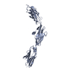 2nziS S: Starting model for refinement |
|---|---|
| Similar structure data |
-Links
 PDBj
PDBj


- Assembly
| Deposited unit | 
| ||||||||||||
|---|---|---|---|---|---|---|---|---|---|---|---|---|---|
| 1 | 
| ||||||||||||
| 2 | 
| ||||||||||||
| Unit cell |
|
-Components
| #1: Protein | / Connectin / Rhabdomyosarcoma antigen MU-RMS-40.14 Mass: 33543.184 Da / Num. of mol.: 2 Source method: isolated from a genetically manipulated source Source: (gene. exp.) Homo sapiens (human) / Gene: TTN / Production host:  Escherichia coli (E. coli) Escherichia coli (E. coli)References: UniProt: Q8WZ42, non-specific serine/threonine protein kinase#2: Chemical | Ethylene glycol  Mass: 62.068 Da / Num. of mol.: 2 / Source method: obtained synthetically / Formula: C2H6O2 Mass: 62.068 Da / Num. of mol.: 2 / Source method: obtained synthetically / Formula: C2H6O2#3: Water | ChemComp-HOH / | Water Mass: 18.015 Da / Num. of mol.: 132 / Source method: isolated from a natural source / Formula: H2O Mass: 18.015 Da / Num. of mol.: 132 / Source method: isolated from a natural source / Formula: H2OHas ligand of interest | N | |
|---|
-Experimental details
-Experiment
| Experiment | Method: X-RAY DIFFRACTION / Number of used crystals: 1 |
|---|
- Sample preparation
| Crystal | Density Matthews: 2.47 Å3/Da / Density % sol: 50.28 % |
|---|---|
| Crystal grow | Temperature: 277.15 K / Method: vapor diffusion, sitting drop Details: 5% [v/v] polyethylene glycol 400 and 40% [v/v] 2-methyl-2,4-pentanediol (MPD) |
-Data collection
| Diffraction | Mean temperature: 293.15 K / Serial crystal experiment: N |
|---|---|
| Diffraction source | Source: SYNCHROTRON / Site: Diamond / Beamline: I04 / Wavelength: 0.9725 Å |
| Detector | Type: DECTRIS PILATUS 6M / Detector: PIXEL / Date: Mar 7, 2015 |
| Radiation | Protocol: SINGLE WAVELENGTH / Monochromatic (M) / Laue (L): M / Scattering type: x-ray |
| Radiation wavelength | Wavelength: 0.9725 Å / Relative weight: 1 |
| Reflection | Resolution: 2.3→29.5 Å / Num. obs: 27975 / % possible obs: 97.95 % / Redundancy: 3.6 % / Biso Wilson estimate: 45.69 Å2 / CC1/2: 0.996 / Rrim(I) all: 0.121 / Net I/σ(I): 9.27 |
| Reflection shell | Resolution: 2.3→2.35 Å / Redundancy: 3.6 % / Mean I/σ(I) obs: 1.02 / Num. unique obs: 2819 / CC1/2: 0.492 / % possible all: 97.1 |
- Processing
| Software |
| ||||||||||||||||||||||||||||||||||||||||||||||||||||||||
|---|---|---|---|---|---|---|---|---|---|---|---|---|---|---|---|---|---|---|---|---|---|---|---|---|---|---|---|---|---|---|---|---|---|---|---|---|---|---|---|---|---|---|---|---|---|---|---|---|---|---|---|---|---|---|---|---|---|
| Refinement | Method to determine structure: MOLECULAR REPLACEMENT Starting model: 2NZI Resolution: 2.3→29.5 Å / SU ML: 0.3396 / Cross valid method: FREE R-VALUE / σ(F): 1.96 / Phase error: 32.6361 Stereochemistry target values: GeoStd + Monomer Library + CDL v1.2
| ||||||||||||||||||||||||||||||||||||||||||||||||||||||||
| Solvent computation | Shrinkage radii: 0.9 Å / VDW probe radii: 1.11 Å / Solvent model: FLAT BULK SOLVENT MODEL | ||||||||||||||||||||||||||||||||||||||||||||||||||||||||
| Displacement parameters | Biso mean: 63.34 Å2 | ||||||||||||||||||||||||||||||||||||||||||||||||||||||||
| Refinement step | Cycle: LAST / Resolution: 2.3→29.5 Å
| ||||||||||||||||||||||||||||||||||||||||||||||||||||||||
| Refine LS restraints |
| ||||||||||||||||||||||||||||||||||||||||||||||||||||||||
| LS refinement shell |
|
