 Movie
Movie Controller


+ Open data

- Basic information
| Entry | Database: PDB / ID: 8blo | |||||||||
|---|---|---|---|---|---|---|---|---|---|---|
| Title | Human Urea Transporter UT-A (N-Terminal Domain Model) | |||||||||
 Components Components | Urea transporter 2 | |||||||||
 Keywords Keywords | TRANSPORT PROTEIN / SLC14A2 / UT2 / UT-A / Urea Transporter / Inhibitor / Solute Carrier | |||||||||
| Function / homology |  Function and homology information Function and homology informationurea transport / Transport of bile salts and organic acids, metal ions and amine compounds / urea transmembrane transporter activity / urea transmembrane transport / cell adhesion molecule binding / transmembrane transport / apical plasma membrane / membrane / plasma membraneSimilarity search - Function | |||||||||
| Biological species |  Homo sapiens (human) Homo sapiens (human) | |||||||||
| Method | ELECTRON MICROSCOPY / single particle reconstruction / cryo EM / Resolution: 2.9 Å | |||||||||
 Authors Authors | Chi, G. / Pike, A.C.W. / Maclean, E.M. / Mukhopadhyay, S.M.M. / Bohstedt, T. / Scacioc, A. / Wang, D. / McKinley, G. / Fernandez-Cid, A. / Arrowsmith, C.H. ...Chi, G. / Pike, A.C.W. / Maclean, E.M. / Mukhopadhyay, S.M.M. / Bohstedt, T. / Scacioc, A. / Wang, D. / McKinley, G. / Fernandez-Cid, A. / Arrowsmith, C.H. / Bountra, C. / Edwards, A. / Burgess-Brown, N.A. / van Putte, W. / Duerr, K. | |||||||||
| Funding support |  United Kingdom, European Union, 2items United Kingdom, European Union, 2items
| |||||||||
 Citation Citation | Journal: Sci Adv / Year: 2023 Title: Structural characterization of human urea transporters UT-A and UT-B and their inhibition. Authors: Gamma Chi / Larissa Dietz / Haiping Tang / Matthew Snee / Andreea Scacioc / Dong Wang / Gavin Mckinley / Shubhashish M M Mukhopadhyay / Ashley C W Pike / Rod Chalk / Nicola A Burgess-Brown / ...Authors: Gamma Chi / Larissa Dietz / Haiping Tang / Matthew Snee / Andreea Scacioc / Dong Wang / Gavin Mckinley / Shubhashish M M Mukhopadhyay / Ashley C W Pike / Rod Chalk / Nicola A Burgess-Brown / Jean-Pierre Timmermans / Wouter van Putte / Carol V Robinson / Katharina L Dürr /  Abstract: In this study, we present the structures of human urea transporters UT-A and UT-B to characterize them at molecular level and to detail the mechanism of UT-B inhibition by its selective inhibitor, ...In this study, we present the structures of human urea transporters UT-A and UT-B to characterize them at molecular level and to detail the mechanism of UT-B inhibition by its selective inhibitor, UTB-14. High-resolution structures of both transporters establish the structural basis for the inhibitor's selectivity to UT-B, and the identification of multiple binding sites for the inhibitor will aid with the development of drug lead molecules targeting both transporters. Our study also discovers phospholipids associating with the urea transporters by combining structural observations, native MS, and lipidomics analysis. These insights improve our understanding of urea transporter function at a molecular level and provide a blueprint for a structure-guided design of therapeutics targeting these transporters. | |||||||||
| History |
|
- Structure visualization
| Structure viewer | Molecule: MolmilJmol/JSmol |
|---|
- Downloads & links
-Download
| PDBx/mmCIF format | 8blo.cif.gz | 256.5 KB | Display | PDBx/mmCIF format |
|---|---|---|---|---|
| PDB format | pdb8blo.ent.gz | 180.3 KB | Display | PDB format |
| PDBx/mmJSON format | 8blo.json.gz | Tree view | PDBx/mmJSON format | |
| Others |  Other downloads Other downloads |
-Validation report
| Arichive directory | https://data.pdbj.org/pub/pdb/validation_reports/bl/8bloftp://data.pdbj.org/pub/pdb/validation_reports/bl/8blo | HTTPS FTP |
|---|
-Related structure data
| Related structure data |  16110MC 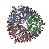 8blpC M: map data used to model this data C: citing same article ( |
|---|---|
| Similar structure data |
-Links
 PDBj
PDBj- Assembly
| Deposited unit | 
|
|---|---|
| 1 |
|
-Components
| #1: Protein | / Solute carrier family 14 member 2 / Urea transporter / kidney Mass: 100124.172 Da / Num. of mol.: 3 Source method: isolated from a genetically manipulated source Details: This model is based on the N-terminal half of the protein UT-A. However, UT-A consists of two similar domains, and the map is sometimes ambiguous whether the structure is of the N-terminal ...Details: This model is based on the N-terminal half of the protein UT-A. However, UT-A consists of two similar domains, and the map is sometimes ambiguous whether the structure is of the N-terminal or C-terminal half. Much of the map is in line with amino acids from its N-terminal domain. Source: (gene. exp.) Homo sapiens (human) / Gene: SLC14A2, HUT2, UT2 / Production host: Homo sapiens (human) / References: UniProt: Q15849#2: Chemical | ChemComp-PLD /   Mass: 875.313 Da / Num. of mol.: 9 / Source method: obtained synthetically / Formula: C50H101NO8P Mass: 875.313 Da / Num. of mol.: 9 / Source method: obtained synthetically / Formula: C50H101NO8P#3: Chemical | ChemComp-LMN /   Mass: 1005.188 Da / Num. of mol.: 15 / Source method: obtained synthetically / Formula: C47H88O22 / Comment: detergent*YM Mass: 1005.188 Da / Num. of mol.: 15 / Source method: obtained synthetically / Formula: C47H88O22 / Comment: detergent*YM#4: Water | ChemComp-HOH / | Water Mass: 18.015 Da / Num. of mol.: 18 / Source method: isolated from a natural source / Formula: H2O Mass: 18.015 Da / Num. of mol.: 18 / Source method: isolated from a natural source / Formula: H2OHas ligand of interest | N | |
|---|
-Experimental details
-Experiment
| Experiment | Method: ELECTRON MICROSCOPY |
|---|---|
| EM experiment | Aggregation state: PARTICLE / 3D reconstruction method: single particle reconstruction |
- Sample preparation
| Component | Name: Trimer-like complex of Human UT-A / Type: COMPLEX / Entity ID: #1 / Source: RECOMBINANT | |||||||||||||||||||||||||
|---|---|---|---|---|---|---|---|---|---|---|---|---|---|---|---|---|---|---|---|---|---|---|---|---|---|---|
| Molecular weight | Value: 0.12 MDa / Experimental value: NO | |||||||||||||||||||||||||
| Source (natural) | Organism: Homo sapiens (human) | |||||||||||||||||||||||||
| Source (recombinant) | Organism: Homo sapiens (human) | |||||||||||||||||||||||||
| Buffer solution | pH: 7.5 | |||||||||||||||||||||||||
| Buffer component |
| |||||||||||||||||||||||||
| Specimen | Embedding applied: NO / Shadowing applied: NO / Staining applied: NO / Vitrification applied: YES | |||||||||||||||||||||||||
| Specimen support | Grid type: Homemade | |||||||||||||||||||||||||
| Vitrification | Cryogen name: ETHANE |
- Electron microscopy imaging
| Experimental equipment |  Model: Titan Krios / Image courtesy: FEI Company |
|---|---|
| Microscopy | Model: TFS KRIOS |
| Electron gun | Electron source: FIELD EMISSION GUN / Accelerating voltage: 300 kV / Illumination mode: FLOOD BEAM |
| Electron lens | Mode: BRIGHT FIELDBright-field microscopy / Nominal defocus max: 2800 nm / Nominal defocus min: 1000 nm |
| Image recording | Electron dose: 52.63 e/Å2 / Film or detector model: GATAN K2 SUMMIT (4k x 4k) |
- Processing
| Software | Name: PHENIX / Version: 1.20.1_4487: / Classification: refinement |
|---|---|
| EM software | Name: cryoSPARC / Category: 3D reconstruction |
| CTF correction | Type: PHASE FLIPPING AND AMPLITUDE CORRECTION |
| Symmetry | Point symmetry: C3 (3 fold cyclic) |
| 3D reconstruction | Resolution: 2.9 Å / Resolution method: FSC 0.143 CUT-OFF / Num. of particles: 180448 / Symmetry type: POINT |
