 Movie
Movie Controller


+ Open data

- Basic information
| Entry |  | |||||||||
|---|---|---|---|---|---|---|---|---|---|---|
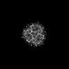
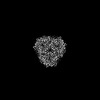
| Title | Human Urea Transporter UT-A (N-Terminal Domain Model) | |||||||||
 Map data Map data | ||||||||||
 Sample Sample |
| |||||||||
 Keywords Keywords | SLC14A2 / UT2 / UT-A /  Urea Transporter / Inhibitor / Solute Carrier / TRANSPORT PROTEIN Urea Transporter / Inhibitor / Solute Carrier / TRANSPORT PROTEIN | |||||||||
| Function / homology |  Function and homology information Function and homology informationurea transport / Transport of bile salts and organic acids, metal ions and amine compounds / urea transmembrane transporter activity / urea transmembrane transport / cell adhesion molecule binding / transmembrane transport / apical plasma membrane / membrane / plasma membraneSimilarity search - Function | |||||||||
| Biological species |  Homo sapiens (human) Homo sapiens (human) | |||||||||
| Method | single particle reconstruction / cryo EM / Resolution: 2.9 Å | |||||||||
 Authors Authors | Chi G / Pike ACW / Maclean EM / Mukhopadhyay SMM / Bohstedt T / Scacioc A / Wang D / McKinley G / Fernandez-Cid A / Arrowsmith CH ...Chi G / Pike ACW / Maclean EM / Mukhopadhyay SMM / Bohstedt T / Scacioc A / Wang D / McKinley G / Fernandez-Cid A / Arrowsmith CH / Bountra C / Edwards A / Burgess-Brown NA / van Putte W / Duerr K | |||||||||
| Funding support |  United Kingdom, European Union, 2 items United Kingdom, European Union, 2 items
| |||||||||
 Citation Citation | Journal: Sci Adv / Year: 2023 Title: Structural characterization of human urea transporters UT-A and UT-B and their inhibition. Authors: Gamma Chi / Larissa Dietz / Haiping Tang / Matthew Snee / Andreea Scacioc / Dong Wang / Gavin Mckinley / Shubhashish M M Mukhopadhyay / Ashley C W Pike / Rod Chalk / Nicola A Burgess-Brown / ...Authors: Gamma Chi / Larissa Dietz / Haiping Tang / Matthew Snee / Andreea Scacioc / Dong Wang / Gavin Mckinley / Shubhashish M M Mukhopadhyay / Ashley C W Pike / Rod Chalk / Nicola A Burgess-Brown / Jean-Pierre Timmermans / Wouter van Putte / Carol V Robinson / Katharina L Dürr /  Abstract: In this study, we present the structures of human urea transporters UT-A and UT-B to characterize them at molecular level and to detail the mechanism of UT-B inhibition by its selective inhibitor, ...In this study, we present the structures of human urea transporters UT-A and UT-B to characterize them at molecular level and to detail the mechanism of UT-B inhibition by its selective inhibitor, UTB-14. High-resolution structures of both transporters establish the structural basis for the inhibitor's selectivity to UT-B, and the identification of multiple binding sites for the inhibitor will aid with the development of drug lead molecules targeting both transporters. Our study also discovers phospholipids associating with the urea transporters by combining structural observations, native MS, and lipidomics analysis. These insights improve our understanding of urea transporter function at a molecular level and provide a blueprint for a structure-guided design of therapeutics targeting these transporters. | |||||||||
| History |
|
- Structure visualization
| Supplemental images |
|---|

- Downloads & links
-EMDB archive
| Map data | emd_16110.map.gz | 97 MB | EMDB map data format | |
|---|---|---|---|---|
| Header (meta data) | emd-16110-v30.xmlemd-16110.xml | 18 KB 18 KB | Display Display | EMDB header |
| FSC (resolution estimation) | emd_16110_fsc.xml | 15.3 KB | Display | FSC data file |
| Images | 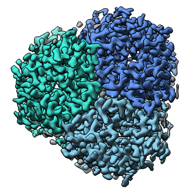 emd_16110.png emd_16110.png | 216.7 KB | ||
| Masks | emd_16110_msk_1.map | 103 MB | Mask map | |
| Filedesc metadata | emd-16110.cif.gz | 6.7 KB | ||
| Others | emd_16110_half_map_1.map.gzemd_16110_half_map_2.map.gz | 95.5 MB 95.5 MB | ||
| Archive directory |  http://ftp.pdbj.org/pub/emdb/structures/EMD-16110ftp://ftp.pdbj.org/pub/emdb/structures/EMD-16110 http://ftp.pdbj.org/pub/emdb/structures/EMD-16110ftp://ftp.pdbj.org/pub/emdb/structures/EMD-16110 | HTTPS FTP |
-Related structure data
| Related structure data |  8bloMC  8blpC M: atomic model generated by this map C: citing same article ( |
|---|---|
| Similar structure data |
-Links
| EMDB pages | EMDB (EBI/PDBe) / EMDataResource |
|---|
-Map
| File | Download / File: emd_16110.map.gz / Format: CCP4 / Size: 103 MB / Type: IMAGE STORED AS FLOATING POINT NUMBER (4 BYTES) | ||||||||||||||||||||||||||||||||||||
|---|---|---|---|---|---|---|---|---|---|---|---|---|---|---|---|---|---|---|---|---|---|---|---|---|---|---|---|---|---|---|---|---|---|---|---|---|---|
| Projections & slices | Image control
Images are generated by Spider. | ||||||||||||||||||||||||||||||||||||
| Voxel size | X=Y=Z: 0.82 Å | ||||||||||||||||||||||||||||||||||||
| Density |
| ||||||||||||||||||||||||||||||||||||
| Symmetry | Space group: 1 | ||||||||||||||||||||||||||||||||||||
| Details | EMDB XML:
|
 Z (Sec.)
 Y (Row.)
 X (Col.)


-Supplemental data
-Mask #1
| File | emd_16110_msk_1.map | ||||||||||||
|---|---|---|---|---|---|---|---|---|---|---|---|---|---|
| Projections & Slices |
| ||||||||||||
| Density Histograms |


-Half map: #1
| File | emd_16110_half_map_1.map | ||||||||||||
|---|---|---|---|---|---|---|---|---|---|---|---|---|---|
| Projections & Slices |
| ||||||||||||
| Density Histograms |


-Half map: #2
| File | emd_16110_half_map_2.map | ||||||||||||
|---|---|---|---|---|---|---|---|---|---|---|---|---|---|
| Projections & Slices |
| ||||||||||||
| Density Histograms |


- Sample components
-Entire : Trimer-like complex of Human UT-A
| Entire | Name: Trimer-like complex of Human UT-A |
|---|---|
| Components |
|
-Supramolecule #1: Trimer-like complex of Human UT-A
| Supramolecule | Name: Trimer-like complex of Human UT-A / type: complex / ID: 1 / Parent: 0 / Macromolecule list: #1 |
|---|---|
| Source (natural) | Organism: Homo sapiens (human) |
| Molecular weight | Theoretical: 120 KDa |
-Macromolecule #1: Urea transporter 2
| Macromolecule | Name: Urea transporter 2 / type: protein_or_peptide / ID: 1 Details: This model is based on the N-terminal half of the protein UT-A. However, UT-A consists of two similar domains, and the map is sometimes ambiguous whether the structure is of the N-terminal ...Details: This model is based on the N-terminal half of the protein UT-A. However, UT-A consists of two similar domains, and the map is sometimes ambiguous whether the structure is of the N-terminal or C-terminal half. Much of the map is in line with amino acids from its N-terminal domain. Number of copies: 3 / Enantiomer: LEVO |
|---|---|
| Source (natural) | Organism: Homo sapiens (human) |
| Molecular weight | Theoretical: 100.124172 KDa |
| Recombinant expression | Organism: Homo sapiens (human) |
| Sequence | String: MSDPHSSPLL PEPLSSRYKL YEAEFTSPSW PSTSPDTHPA LPLLEMPEEK DLRSSNEDSH IVKIEKLNER SKRKDDGVAH RDSAGQRCI CLSKAVGYLT GDMKEYRIWL KDKHLALQFI DWVLRGTAQV MFINNPLSGL IIFIGLLIQN PWWTITGGLG T VVSTLTAL ...String: MSDPHSSPLL PEPLSSRYKL YEAEFTSPSW PSTSPDTHPA LPLLEMPEEK DLRSSNEDSH IVKIEKLNER SKRKDDGVAH RDSAGQRCI CLSKAVGYLT GDMKEYRIWL KDKHLALQFI DWVLRGTAQV MFINNPLSGL IIFIGLLIQN PWWTITGGLG T VVSTLTAL ALGQDRSAIA SGLHGYNGML VGLLMAVFSE KLDYYWWLLF PVTFTAMSCP VLSSALNSIF SKWDLPVFTL PF NIAVTLY LAATGHYNLF FPTTLVEPVS SVPNITWTEM EMPLLLQAIP VGVGQVYGCD NPWTGGVFLV ALFISSPLIC LHA AIGSIV GLLAALSVAT PFETIYTGLW SYNCVLSCIA IGGMFYALTW QTHLLALICA LFCAYMEAAI SNIMSVVGVP PGTW AFCLA TIIFLLLTTN NPAIFRLPLS KVTYPEANRI YYLTVKSGEE EKAPSGGGGE HPPTAGPKVE EGSEAVLSKH RSVFH IEWS SIRRRSKVFG KGEHQERQNK DPFPYRYRKP TVELLDLDTM EESSEIKVET NISKTSWIRS SMAASGKRVS KALSYI TGE MKECGEGLKD KSPVFQFFDW VLRGTSQVMF VNNPLSGILI ILGLFIQNPW WAISGCLGTI MSTLTALILS QDKSAIA AG FHGYNGVLVG LLMAVFSDKG DYYWWLLLPV IIMSMSCPIL SSALGTIFSK WDLPVFTLPF NITVTLYLAA TGHYNLFF P TTLLQPASAM PNITWSEVQV PLLLRAIPVG IGQVYGCDNP WTGGIFLIAL FISSPLICLH AAIGSTMGML AALTIATPF DSIYFGLCGF NSTLACIAIG GMFYVITWQT HLLAIACALF AAYLGAALAN MLSVFGLPPC TWPFCLSALT FLLLTTNNPA IYKLPLSKV TYPEANRIYY LSQEAENLYF Q UniProtKB: Urea transporter 2 |
-Macromolecule #2: di-heneicosanoyl phosphatidyl choline
| Macromolecule | Name: di-heneicosanoyl phosphatidyl choline / type: ligand / ID: 2 / Number of copies: 9 / Formula: PLD |
|---|---|
| Molecular weight | Theoretical: 875.313 Da |
| Chemical component information |  ChemComp-PLD: |
-Macromolecule #3: Lauryl Maltose Neopentyl Glycol
| Macromolecule | Name: Lauryl Maltose Neopentyl Glycol / type: ligand / ID: 3 / Number of copies: 15 / Formula: LMN |
|---|---|
| Molecular weight | Theoretical: 1.005188 KDa |
| Chemical component information |  ChemComp-AV0: |
-Macromolecule #4: water
| Macromolecule | Name: water / type: ligand / ID: 4 / Number of copies: 18 / Formula: HOH |
|---|---|
| Molecular weight | Theoretical: 18.015 Da |
| Chemical component information |  ChemComp-HOH: |
-Experimental details
-Structure determination
| Method | cryo EM |
|---|---|
 Processing Processing | single particle reconstruction |
| Aggregation state | particle |
-Sample preparation
| Buffer | pH: 7.5 Component:
| |||||||||||||||
|---|---|---|---|---|---|---|---|---|---|---|---|---|---|---|---|---|
| Grid | Model: Homemade / Support film - Material: GRAPHENE | |||||||||||||||
| Vitrification | Cryogen name: ETHANE |
- Electron microscopy
| Microscope | TFS KRIOS |
|---|---|
| Electron beam | Acceleration voltage: 300 kV / Electron source: FIELD EMISSION GUN |
| Electron optics | Illumination mode: FLOOD BEAM / Imaging mode: BRIGHT FIELDBright-field microscopy / Nominal defocus max: 2.8000000000000003 µm / Nominal defocus min: 1.0 µm |
| Image recording | Film or detector model: GATAN K2 SUMMIT (4k x 4k) / Average electron dose: 52.63 e/Å2 |
| Experimental equipment |  Model: Titan Krios / Image courtesy: FEI Company |
-Image processing
| Startup model | Type of model: NONE |
|---|---|
| Initial angle assignment | Type: MAXIMUM LIKELIHOOD |
| Final angle assignment | Type: MAXIMUM LIKELIHOOD |
| Final reconstruction | Applied symmetry - Point group: C3 (3 fold cyclic) / Resolution.type: BY AUTHOR / Resolution: 2.9 Å / Resolution method: FSC 0.143 CUT-OFF / Software - Name: cryoSPARC / Number images used: 180448 |
| FSC plot (resolution estimation) |  |
