 Movie
Movie Controller


+ Open data

- Basic information
| Entry | Database: PDB / ID: 8b8b | |||||||||
|---|---|---|---|---|---|---|---|---|---|---|
| Title | Multimerization domain of Munia virus 1 phosphoprotein | |||||||||
 Components Components | Munia Bornavirus 1 phosphoprotein | |||||||||
 Keywords Keywords |  VIRAL PROTEIN / phosphoprotein / RNA polymerase cofactor VIRAL PROTEIN / phosphoprotein / RNA polymerase cofactor | |||||||||
| Function / homology | NITRATE ION Function and homology information Function and homology information | |||||||||
| Biological species |  Munia Bornavirus 1 Munia Bornavirus 1 | |||||||||
| Method | X-RAY DIFFRACTION / SYNCHROTRON / MOLECULAR REPLACEMENT / molecular replacement / Resolution: 2.15 Å | |||||||||
 Authors Authors | Chenavier, F. / Tarbouriech, N. / Bourhis, J.M. / Tomonaga, K. / Horie, M. / Crepin, T. | |||||||||
| Funding support |  France, France,  Japan, 2items Japan, 2items
| |||||||||
 Citation Citation | Journal: Viruses / Year: 2022 Title: Borna Disease Virus 1 Phosphoprotein Forms a Tetramer and Interacts with Host Factors Involved in DNA Double-Strand Break Repair and mRNA Processing. Authors: Tarbouriech, N. / Chenavier, F. / Kawasaki, J. / Bachiri, K. / Bourhis, J.M. / Legrand, P. / Freslon, L.L. / Laurent, E.M.N. / Suberbielle, E. / Ruigrok, R.W.H. / Tomonaga, K. / Gonzalez- ...Authors: Tarbouriech, N. / Chenavier, F. / Kawasaki, J. / Bachiri, K. / Bourhis, J.M. / Legrand, P. / Freslon, L.L. / Laurent, E.M.N. / Suberbielle, E. / Ruigrok, R.W.H. / Tomonaga, K. / Gonzalez-Dunia, D. / Horie, M. / Coyaud, E. / Crepin, T. | |||||||||
| History |
|
- Structure visualization
| Structure viewer | Molecule: MolmilJmol/JSmol |
|---|
- Downloads & links
-Download
| PDBx/mmCIF format | 8b8b.cif.gz | 88.5 KB | Display | PDBx/mmCIF format |
|---|---|---|---|---|
| PDB format | pdb8b8b.ent.gz | 67.1 KB | Display | PDB format |
| PDBx/mmJSON format | 8b8b.json.gz | Tree view | PDBx/mmJSON format | |
| Others |  Other downloads Other downloads |
-Validation report
| Arichive directory | https://data.pdbj.org/pub/pdb/validation_reports/b8/8b8bftp://data.pdbj.org/pub/pdb/validation_reports/b8/8b8b | HTTPS FTP |
|---|
-Related structure data
| Related structure data | 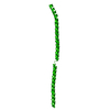 8b8aC 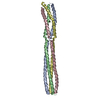 8b8dC C: citing same article ( |
|---|---|
| Similar structure data | |
| Other databases |
|
-Links
 PDBj
PDBj- Assembly
| Deposited unit | 
| ||||||||
|---|---|---|---|---|---|---|---|---|---|
| 1 |
| ||||||||
| Unit cell |
|
-Components
| #1: Protein | Mass: 12342.133 Da / Num. of mol.: 4 Source method: isolated from a genetically manipulated source Source: (gene. exp.) Munia Bornavirus 1 / Plasmid: petM11 / Production host:  Escherichia coli BL21 (bacteria) / Variant (production host): RIL Escherichia coli BL21 (bacteria) / Variant (production host): RIL#2: Chemical | Nitrate  Mass: 62.005 Da / Num. of mol.: 2 / Source method: obtained synthetically / Formula: NO3 Mass: 62.005 Da / Num. of mol.: 2 / Source method: obtained synthetically / Formula: NO3#3: Water | ChemComp-HOH / | Water Mass: 18.015 Da / Num. of mol.: 109 / Source method: isolated from a natural source / Formula: H2O Mass: 18.015 Da / Num. of mol.: 109 / Source method: isolated from a natural source / Formula: H2OHas ligand of interest | N | |
|---|
-Experimental details
-Experiment
| Experiment | Method: X-RAY DIFFRACTION / Number of used crystals: 1 |
|---|
- Sample preparation
| Crystal | Density Matthews: 2.19 Å3/Da / Density % sol: 43.91 % |
|---|---|
| Crystal grow | Temperature: 293 K / Method: vapor diffusion, hanging drop / Details: 15-18 % PEG 3350, 150 mM Mg(NO3)2, 10 mM phenol |
-Data collection
| Diffraction | Mean temperature: 100 K / Serial crystal experiment: N |
|---|---|
| Diffraction source | Source: SYNCHROTRON / Site: SOLEIL / Beamline: PROXIMA 1 / Wavelength: 0.978565 Å |
| Detector | Type: DECTRIS EIGER X 16M / Detector: PIXEL / Date: Apr 8, 2021 |
| Radiation | Protocol: SINGLE WAVELENGTH / Monochromatic (M) / Laue (L): M / Scattering type: x-ray |
| Radiation wavelength | Wavelength: 0.978565 Å / Relative weight: 1 |
| Reflection | Resolution: 2.15→49.34 Å / Num. obs: 23663 / % possible obs: 99.9 % / Redundancy: 5.4 % / CC1/2: 0.998 / Rmerge(I) obs: 0.052 / Rpim(I) all: 0.037 / Net I/σ(I): 15.2 |
| Reflection shell | Resolution: 2.15→2.22 Å / Redundancy: 5.6 % / Rmerge(I) obs: 0.67 / Mean I/σ(I) obs: 2 / Num. unique obs: 2043 / CC1/2: 0.808 / Rpim(I) all: 0.471 / % possible all: 100 |
-Phasing
| Phasing | Method: molecular replacement |
|---|
- Processing
| Software |
| ||||||||||||||||||||||||||||||||||||||||||||||||||||||||||||
|---|---|---|---|---|---|---|---|---|---|---|---|---|---|---|---|---|---|---|---|---|---|---|---|---|---|---|---|---|---|---|---|---|---|---|---|---|---|---|---|---|---|---|---|---|---|---|---|---|---|---|---|---|---|---|---|---|---|---|---|---|---|
| Refinement | Method to determine structure: MOLECULAR REPLACEMENT Starting model: D_1292125760 Resolution: 2.15→49.34 Å / Cor.coef. Fo:Fc: 0.955 / Cor.coef. Fo:Fc free: 0.893 / SU B: 7.697 / SU ML: 0.195 / Cross valid method: THROUGHOUT / σ(F): 0 / ESU R: 0.262 / ESU R Free: 0.254 / Stereochemistry target values: MAXIMUM LIKELIHOOD Details: HYDROGENS HAVE BEEN ADDED IN THE RIDING POSITIONS U VALUES : REFINED INDIVIDUALLY
| ||||||||||||||||||||||||||||||||||||||||||||||||||||||||||||
| Solvent computation | Ion probe radii: 0.8 Å / Shrinkage radii: 0.8 Å / VDW probe radii: 1.2 Å / Solvent model: MASK | ||||||||||||||||||||||||||||||||||||||||||||||||||||||||||||
| Displacement parameters | Biso max: 135.76 Å2 / Biso mean: 54.702 Å2 / Biso min: 22.98 Å2
| ||||||||||||||||||||||||||||||||||||||||||||||||||||||||||||
| Refinement step | Cycle: final / Resolution: 2.15→49.34 Å
| ||||||||||||||||||||||||||||||||||||||||||||||||||||||||||||
| Refine LS restraints |
| ||||||||||||||||||||||||||||||||||||||||||||||||||||||||||||
| LS refinement shell | Resolution: 2.15→2.206 Å / Rfactor Rfree error: 0 / Total num. of bins used: 20
|
