 Movie
Movie Controller


+ Open data

- Basic information
| Entry | Database: PDB / ID: 8a0e | |||||||||
|---|---|---|---|---|---|---|---|---|---|---|
| Title | CryoEM structure of DHS-eIF5A1 complex | |||||||||
 Components Components |
| |||||||||
 Keywords Keywords |  TRANSFERASE / Hypusination / posttranslational modification TRANSFERASE / Hypusination / posttranslational modification | |||||||||
| Function / homology |  Function and homology informationdeoxyhypusine synthase / deoxyhypusine synthase activity / Hypusine synthesis from eIF5A-lysine / peptidyl-lysine modification to peptidyl-hypusine / spermidine metabolic process / spermidine catabolic process / positive regulation of translational termination / positive regulation of translational elongation / translation elongation factor activity / positive regulation of T cell proliferation ...deoxyhypusine synthase / deoxyhypusine synthase activity / Hypusine synthesis from eIF5A-lysine / peptidyl-lysine modification to peptidyl-hypusine / spermidine metabolic process / spermidine catabolic process / positive regulation of translational termination / positive regulation of translational elongation / translation elongation factor activity / positive regulation of T cell proliferation / translation initiation factor activity / ribosome binding / glucose homeostasis / translation / positive regulation of cell population proliferation / endoplasmic reticulum membrane / RNA binding / identical protein binding / cytosol / cytoplasm Function and homology informationdeoxyhypusine synthase / deoxyhypusine synthase activity / Hypusine synthesis from eIF5A-lysine / peptidyl-lysine modification to peptidyl-hypusine / spermidine metabolic process / spermidine catabolic process / positive regulation of translational termination / positive regulation of translational elongation / translation elongation factor activity / positive regulation of T cell proliferation ...deoxyhypusine synthase / deoxyhypusine synthase activity / Hypusine synthesis from eIF5A-lysine / peptidyl-lysine modification to peptidyl-hypusine / spermidine metabolic process / spermidine catabolic process / positive regulation of translational termination / positive regulation of translational elongation / translation elongation factor activity / positive regulation of T cell proliferation / translation initiation factor activity / ribosome binding / glucose homeostasis / translation / positive regulation of cell population proliferation / endoplasmic reticulum membrane / RNA binding / identical protein binding / cytosol / cytoplasmSimilarity search - Function | |||||||||
| Biological species |  Homo sapiens (human) Homo sapiens (human) | |||||||||
| Method | ELECTRON MICROSCOPY / single particle reconstruction / cryo EM / Resolution: 2.8 Å | |||||||||
 Authors Authors | Wator, E. / Wilk, P. / Biela, A.P. / Rawski, M. / Grudnik, P. | |||||||||
| Funding support |  Poland, 2items Poland, 2items
| |||||||||
 Citation Citation | Journal: Nat Commun / Year: 2023 Title: Cryo-EM structure of human eIF5A-DHS complex reveals the molecular basis of hypusination-associated neurodegenerative disorders. Authors: Elżbieta Wątor / Piotr Wilk / Artur Biela / Michał Rawski / Krzysztof M Zak / Wieland Steinchen / Gert Bange / Sebastian Glatt / Przemysław Grudnik /  Abstract: Hypusination is a unique post-translational modification of the eukaryotic translation factor 5A (eIF5A) that is essential for overcoming ribosome stalling at polyproline sequence stretches. The ...Hypusination is a unique post-translational modification of the eukaryotic translation factor 5A (eIF5A) that is essential for overcoming ribosome stalling at polyproline sequence stretches. The initial step of hypusination, the formation of deoxyhypusine, is catalyzed by deoxyhypusine synthase (DHS), however, the molecular details of the DHS-mediated reaction remained elusive. Recently, patient-derived variants of DHS and eIF5A have been linked to rare neurodevelopmental disorders. Here, we present the cryo-EM structure of the human eIF5A-DHS complex at 2.8 Å resolution and a crystal structure of DHS trapped in the key reaction transition state. Furthermore, we show that disease-associated DHS variants influence the complex formation and hypusination efficiency. Hence, our work dissects the molecular details of the deoxyhypusine synthesis reaction and reveals how clinically-relevant mutations affect this crucial cellular process. | |||||||||
| History |
|
- Structure visualization
| Structure viewer | Molecule: MolmilJmol/JSmol |
|---|
- Downloads & links
-Download
| PDBx/mmCIF format | 8a0e.cif.gz | 265.7 KB | Display | PDBx/mmCIF format |
|---|---|---|---|---|
| PDB format | pdb8a0e.ent.gz | 219.6 KB | Display | PDB format |
| PDBx/mmJSON format | 8a0e.json.gz | Tree view | PDBx/mmJSON format | |
| Others |  Other downloads Other downloads |
-Validation report
| Arichive directory | https://data.pdbj.org/pub/pdb/validation_reports/a0/8a0eftp://data.pdbj.org/pub/pdb/validation_reports/a0/8a0e | HTTPS FTP |
|---|
-Related structure data
| Related structure data |  15052MC 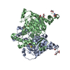 7a6sC  7a6tC 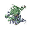 8a0fC  8a0gC C: citing same article ( M: map data used to model this data |
|---|---|
| Similar structure data |

-Links
 PDBj
PDBj


- Assembly
| Deposited unit | 
|
|---|---|
| 1 |
|
-Components
| #1: Protein | / DHS Mass: 41098.461 Da / Num. of mol.: 2 / Mutation: K329A Source method: isolated from a genetically manipulated source Source: (gene. exp.) Homo sapiens (human) / Gene: DHPS, DS / Production host:  Escherichia coli (E. coli) / References: UniProt: P49366, deoxyhypusine synthase Escherichia coli (E. coli) / References: UniProt: P49366, deoxyhypusine synthase#2: Protein | / DHSMass: 41130.527 Da / Num. of mol.: 2 Source method: isolated from a genetically manipulated source Source: (gene. exp.) Homo sapiens (human) / Gene: DHPS, DS / Production host: Escherichia coli (E. coli) / References: UniProt: P49366, deoxyhypusine synthase#3: Protein | | Mass: 16998.395 Da / Num. of mol.: 1 Source method: isolated from a genetically manipulated source Source: (gene. exp.) Homo sapiens (human) / Gene: EIF5A / Production host: Escherichia coli (E. coli) / References: UniProt: A0A2Y9EFS4#4: Chemical | ChemComp-NAD / Nicotinamide adenine dinucleotide  Mass: 663.425 Da / Num. of mol.: 4 / Source method: obtained synthetically / Formula: C21H27N7O14P2 / Comment: NAD*YM Mass: 663.425 Da / Num. of mol.: 4 / Source method: obtained synthetically / Formula: C21H27N7O14P2 / Comment: NAD*YM#5: Chemical | Spermidine  Mass: 145.246 Da / Num. of mol.: 3 / Source method: obtained synthetically / Formula: C7H19N3 Mass: 145.246 Da / Num. of mol.: 3 / Source method: obtained synthetically / Formula: C7H19N3Has ligand of interest | N | |
|---|
-Experimental details
-Experiment
| Experiment | Method: ELECTRON MICROSCOPY |
|---|---|
| EM experiment | Aggregation state: PARTICLE / 3D reconstruction method: single particle reconstruction |
- Sample preparation
| Component | Name: Complex of human DHS-eIF5A / Type: COMPLEX / Details: monomer of eIF5A bound to homotetramer DHS. / Entity ID: #1-#3 / Source: RECOMBINANT | |||||||||||||||
|---|---|---|---|---|---|---|---|---|---|---|---|---|---|---|---|---|
| Molecular weight | Value: 0.181687 MDa / Experimental value: NO | |||||||||||||||
| Source (natural) | Organism: Homo sapiens (human) | |||||||||||||||
| Source (recombinant) | Organism: Escherichia coli (E. coli) | |||||||||||||||
| Buffer solution | pH: 9.3 | |||||||||||||||
| Buffer component |
| |||||||||||||||
| Specimen | Conc.: 0.12 mg/ml / Embedding applied: NO / Shadowing applied: NO / Staining applied: NO / Vitrification applied: YES | |||||||||||||||
| Specimen support | Grid material: COPPER / Grid mesh size: 400 divisions/in. / Grid type: Quantifoil R2/2 | |||||||||||||||
| Vitrification | Instrument: FEI VITROBOT MARK IV / Cryogen name: ETHANE / Humidity: 100 % / Chamber temperature: 278 K |
- Electron microscopy imaging
| Experimental equipment |  Model: Titan Krios / Image courtesy: FEI Company |
|---|---|
| Microscopy | Model: FEI TITAN KRIOS |
| Electron gun | Electron source: FIELD EMISSION GUN / Accelerating voltage: 300 kV / Illumination mode: FLOOD BEAM |
| Electron lens | Mode: BRIGHT FIELDBright-field microscopy / Nominal defocus max: 3500 nm / Nominal defocus min: 500 nm / Cs: 2.7 mm |
| Specimen holder | Cryogen: NITROGEN |
| Image recording | Electron dose: 40 e/Å2 / Film or detector model: GATAN K3 (6k x 4k) |
- Processing
| EM software |
| |||||||||||||||||||||||||||
|---|---|---|---|---|---|---|---|---|---|---|---|---|---|---|---|---|---|---|---|---|---|---|---|---|---|---|---|---|
| Image processing | Details: selected images were normalized and low-pass filtered | |||||||||||||||||||||||||||
| CTF correction | Type: PHASE FLIPPING AND AMPLITUDE CORRECTION | |||||||||||||||||||||||||||
| Particle selection | Num. of particles selected: 1277576 / Details: autopicked particles | |||||||||||||||||||||||||||
| Symmetry | Point symmetry: C1 (asymmetric) | |||||||||||||||||||||||||||
| 3D reconstruction | Resolution: 2.8 Å / Resolution method: FSC 0.143 CUT-OFF / Num. of particles: 490582 / Algorithm: FOURIER SPACE / Num. of class averages: 1 / Symmetry type: POINT | |||||||||||||||||||||||||||
| Atomic model building | Protocol: RIGID BODY FIT / Space: REAL | |||||||||||||||||||||||||||
| Atomic model building | PDB-ID: 6XXJ |
