 Movie
Movie Controller

[English] 日本語
 Yorodumi
- PDB-7zyt: Crystal structure of the I318T pathogenic variant of the human di... -
+ Open data

- Basic information
| Entry | Database: PDB / ID: 7zyt | ||||||||||||
|---|---|---|---|---|---|---|---|---|---|---|---|---|---|
| Title | Crystal structure of the I318T pathogenic variant of the human dihydrolipoamide dehydrogenase | ||||||||||||
 Components Components | Dihydrolipoyl dehydrogenase, mitochondrial Dihydrolipoamide dehydrogenase Dihydrolipoamide dehydrogenase | ||||||||||||
 Keywords Keywords | OXIDOREDUCTASE / lipoamide dehydrogenase / pathogenic mutation / E3 deficiency / alpha-ketoglutarate dehydrogenase complex / 2-oxoglutarate dehydrogenase complex / pyruvate dehydrogenase complex | ||||||||||||
| Function / homology |  Function and homology informationacetyltransferase complex / acrosomal matrix / Glycine degradation / : / dihydrolipoyl dehydrogenase / dihydrolipoyl dehydrogenase activity / oxoglutarate dehydrogenase complex / acetyl-CoA biosynthetic process from pyruvate / pyruvate dehydrogenase complex / : ...acetyltransferase complex / acrosomal matrix / Glycine degradation / : / dihydrolipoyl dehydrogenase / dihydrolipoyl dehydrogenase activity / oxoglutarate dehydrogenase complex / acetyl-CoA biosynthetic process from pyruvate / pyruvate dehydrogenase complex / : / Lysine catabolism / branched-chain amino acid catabolic process / Citric acid cycle (TCA cycle) / Branched-chain amino acid catabolism / Pyruvate metabolism / Glyoxylate metabolism and glycine degradation / Regulation of pyruvate dehydrogenase (PDH) complex / motile cilium / sperm capacitation / Signaling by Retinoic Acid / mitochondrial electron transport, NADH to ubiquinone / Mitochondrial protein degradation / gastrulation / regulation of membrane potential / flavin adenine dinucleotide binding / mitochondrial matrix / mitochondrion / proteolysis / nucleus Function and homology informationacetyltransferase complex / acrosomal matrix / Glycine degradation / : / dihydrolipoyl dehydrogenase / dihydrolipoyl dehydrogenase activity / oxoglutarate dehydrogenase complex / acetyl-CoA biosynthetic process from pyruvate / pyruvate dehydrogenase complex / : ...acetyltransferase complex / acrosomal matrix / Glycine degradation / : / dihydrolipoyl dehydrogenase / dihydrolipoyl dehydrogenase activity / oxoglutarate dehydrogenase complex / acetyl-CoA biosynthetic process from pyruvate / pyruvate dehydrogenase complex / : / Lysine catabolism / branched-chain amino acid catabolic process / Citric acid cycle (TCA cycle) / Branched-chain amino acid catabolism / Pyruvate metabolism / Glyoxylate metabolism and glycine degradation / Regulation of pyruvate dehydrogenase (PDH) complex / motile cilium / sperm capacitation / Signaling by Retinoic Acid / mitochondrial electron transport, NADH to ubiquinone / Mitochondrial protein degradation / gastrulation / regulation of membrane potential / flavin adenine dinucleotide binding / mitochondrial matrix / mitochondrion / proteolysis / nucleusSimilarity search - Function | ||||||||||||
| Biological species |  Homo sapiens (human) Homo sapiens (human) | ||||||||||||
| Method | X-RAY DIFFRACTION / SYNCHROTRON / MOLECULAR REPLACEMENT / molecular replacement / Resolution: 2.892 Å | ||||||||||||
 Authors Authors | Nemes-Nikodem, E. / Szabo, E. / Vass, K.R. / Lennartz, F. / Nagy, B. / Torocsik, B. / Weiss, M.S. / Adam-Vizi, V. / Ambrus, A. | ||||||||||||
| Funding support |  Hungary, European Union, 3items Hungary, European Union, 3items
| ||||||||||||
 Citation Citation | Journal: Int J Mol Sci / Year: 2023 Title: Structural and Biochemical Investigation of Selected Pathogenic Mutants of the Human Dihydrolipoamide Dehydrogenase. Authors: Szabo, E. / Nemes-Nikodem, E. / Vass, K.R. / Zambo, Z. / Zrupko, E. / Torocsik, B. / Ozohanics, O. / Nagy, B. / Ambrus, A. | ||||||||||||
| History |
|
- Structure visualization
| Structure viewer | Molecule: MolmilJmol/JSmol |
|---|
- Downloads & links
-Download
| PDBx/mmCIF format | 7zyt.cif.gz | 191.1 KB | Display | PDBx/mmCIF format |
|---|---|---|---|---|
| PDB format | pdb7zyt.ent.gz | 148.8 KB | Display | PDB format |
| PDBx/mmJSON format | 7zyt.json.gz | Tree view | PDBx/mmJSON format | |
| Others |  Other downloads Other downloads |
-Validation report
| Arichive directory | https://data.pdbj.org/pub/pdb/validation_reports/zy/7zytftp://data.pdbj.org/pub/pdb/validation_reports/zy/7zyt | HTTPS FTP |
|---|
-Related structure data
| Related structure data | 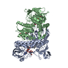 7pscC  6i4qS S: Starting model for refinement C: citing same article ( |
|---|---|
| Similar structure data |
-Links
 PDBj
PDBj


- Assembly
| Deposited unit | 
| ||||||||
|---|---|---|---|---|---|---|---|---|---|
| 1 |
| ||||||||
| Unit cell |
| ||||||||
| Components on special symmetry positions |
|
-Components
| #1: Protein | Dihydrolipoamide dehydrogenase / Dihydrolipoamide dehydrogenase / Glycine cleavage system L protein Mass: 52625.117 Da / Num. of mol.: 2 / Mutation: I318T Source method: isolated from a genetically manipulated source Details: Sequence of the Strep-tag with the linker amino acids: MASWSHPQFEKGALEVLFQGPG Source: (gene. exp.) Homo sapiens (human) / Gene: DLD, GCSL, LAD, PHE3 / Plasmid: pET52b+ / Production host:  Escherichia coli BL21(DE3) (bacteria) / References: UniProt: P09622, dihydrolipoyl dehydrogenase Escherichia coli BL21(DE3) (bacteria) / References: UniProt: P09622, dihydrolipoyl dehydrogenase#2: Chemical | Flavin adenine dinucleotide  Mass: 785.550 Da / Num. of mol.: 2 / Source method: obtained synthetically / Formula: C27H33N9O15P2 / Feature type: SUBJECT OF INVESTIGATION / Comment: FAD*YM Mass: 785.550 Da / Num. of mol.: 2 / Source method: obtained synthetically / Formula: C27H33N9O15P2 / Feature type: SUBJECT OF INVESTIGATION / Comment: FAD*YM#3: Chemical | Bis-tris methane  Mass: 209.240 Da / Num. of mol.: 2 / Source method: obtained synthetically / Formula: C8H19NO5 / Comment: pH buffer*YM Mass: 209.240 Da / Num. of mol.: 2 / Source method: obtained synthetically / Formula: C8H19NO5 / Comment: pH buffer*YM#4: Chemical | ChemComp-EDO / | Ethylene glycol  Mass: 62.068 Da / Num. of mol.: 1 / Source method: obtained synthetically / Formula: C2H6O2 Mass: 62.068 Da / Num. of mol.: 1 / Source method: obtained synthetically / Formula: C2H6O2Has ligand of interest | Y | |
|---|
-Experimental details
-Experiment
| Experiment | Method: X-RAY DIFFRACTION / Number of used crystals: 1 |
|---|
- Sample preparation
| Crystal | Density Matthews: 2.16 Å3/Da / Density % sol: 42.98 % |
|---|---|
| Crystal grow | Temperature: 293 K / Method: vapor diffusion, sitting drop / pH: 6.7 Details: 0.2 M magnesium chloride, 0.1 M BIS-TRIS (pH 6.7), 29 (v/v)% PEG 3350, cryo-protection: 15 and then 30 (v/v)% ethylene glycol in the reservoir |
-Data collection
| Diffraction | Mean temperature: 100 K / Serial crystal experiment: N | ||||||||||||||||||||||||||||||||||||||||||||||||||||||||||||||||||||||||||||||||||||||||||||||||||||
|---|---|---|---|---|---|---|---|---|---|---|---|---|---|---|---|---|---|---|---|---|---|---|---|---|---|---|---|---|---|---|---|---|---|---|---|---|---|---|---|---|---|---|---|---|---|---|---|---|---|---|---|---|---|---|---|---|---|---|---|---|---|---|---|---|---|---|---|---|---|---|---|---|---|---|---|---|---|---|---|---|---|---|---|---|---|---|---|---|---|---|---|---|---|---|---|---|---|---|---|---|---|
| Diffraction source | Source: SYNCHROTRON / Site: BESSY  / Beamline: 14.1 / Wavelength: 0.9184 Å / Beamline: 14.1 / Wavelength: 0.9184 Å | ||||||||||||||||||||||||||||||||||||||||||||||||||||||||||||||||||||||||||||||||||||||||||||||||||||
| Detector | Type: DECTRIS PILATUS3 6M / Detector: PIXEL / Date: Oct 23, 2021 | ||||||||||||||||||||||||||||||||||||||||||||||||||||||||||||||||||||||||||||||||||||||||||||||||||||
| Radiation | Protocol: SINGLE WAVELENGTH / Monochromatic (M) / Laue (L): M / Scattering type: x-ray | ||||||||||||||||||||||||||||||||||||||||||||||||||||||||||||||||||||||||||||||||||||||||||||||||||||
| Radiation wavelength | Wavelength: 0.9184 Å / Relative weight: 1 | ||||||||||||||||||||||||||||||||||||||||||||||||||||||||||||||||||||||||||||||||||||||||||||||||||||
| Reflection | Resolution: 2.88→44.14 Å / Num. obs: 20384 / % possible obs: 98.3 % / Redundancy: 6.439 % / Biso Wilson estimate: 87.275 Å2 / CC1/2: 0.998 / Rmerge(I) obs: 0.182 / Rrim(I) all: 0.198 / Χ2: 0.74 / Net I/σ(I): 8.37 | ||||||||||||||||||||||||||||||||||||||||||||||||||||||||||||||||||||||||||||||||||||||||||||||||||||
| Reflection shell | Diffraction-ID: 1
|
-Phasing
| Phasing | Method: molecular replacement | ||||||
|---|---|---|---|---|---|---|---|
| Phasing MR | R rigid body: 0.604
|
- Processing
| Software |
| ||||||||||||||||||||||||||||||||||||||||||||||||
|---|---|---|---|---|---|---|---|---|---|---|---|---|---|---|---|---|---|---|---|---|---|---|---|---|---|---|---|---|---|---|---|---|---|---|---|---|---|---|---|---|---|---|---|---|---|---|---|---|---|
| Refinement | Method to determine structure: MOLECULAR REPLACEMENT Starting model: 6I4Q Resolution: 2.892→44.14 Å / SU ML: 0.66 / Cross valid method: THROUGHOUT / σ(F): 1.33 / Phase error: 41.83 / Stereochemistry target values: ML Details: refinement was carried out using NCS torsion-angle restraints
| ||||||||||||||||||||||||||||||||||||||||||||||||
| Solvent computation | Shrinkage radii: 0.9 Å / VDW probe radii: 1.11 Å / Solvent model: FLAT BULK SOLVENT MODEL | ||||||||||||||||||||||||||||||||||||||||||||||||
| Displacement parameters | Biso max: 194.76 Å2 / Biso mean: 119.314 Å2 / Biso min: 64.24 Å2 | ||||||||||||||||||||||||||||||||||||||||||||||||
| Refinement step | Cycle: final / Resolution: 2.892→44.14 Å
| ||||||||||||||||||||||||||||||||||||||||||||||||
| LS refinement shell | Refine-ID: X-RAY DIFFRACTION / Rfactor Rfree error: 0
|
