Movie
Movie Controller

[English] 日本語
 Yorodumi
- PDB-7zrv: cryo-EM structure of omicron spike in complex with de novo design... -
+ Open data

- Basic information
| Entry | Database: PDB / ID: 7zrv | ||||||||||||||||||
|---|---|---|---|---|---|---|---|---|---|---|---|---|---|---|---|---|---|---|---|
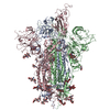
| Title | cryo-EM structure of omicron spike in complex with de novo designed binder, full map | ||||||||||||||||||
 Components Components |
| ||||||||||||||||||
 Keywords Keywords |  ANTIVIRAL PROTEIN / SARS-COV2 / de novo / design binder / spike / RBD / receptor binding domain ANTIVIRAL PROTEIN / SARS-COV2 / de novo / design binder / spike / RBD / receptor binding domain | ||||||||||||||||||
| Function / homology |  Function and homology information Function and homology informationMaturation of spike protein / viral translation / Translation of Structural Proteins / Virion Assembly and Release / host cell surface / host extracellular space / suppression by virus of host tetherin activity / Induction of Cell-Cell Fusion / structural constituent of virion / host cell endoplasmic reticulum-Golgi intermediate compartment membrane ...Maturation of spike protein / viral translation / Translation of Structural Proteins / Virion Assembly and Release / host cell surface / host extracellular space / suppression by virus of host tetherin activity / Induction of Cell-Cell Fusion / structural constituent of virion / host cell endoplasmic reticulum-Golgi intermediate compartment membrane / entry receptor-mediated virion attachment to host cell / receptor-mediated endocytosis of virus by host cell / Attachment and Entry / membrane fusion / positive regulation of viral entry into host cell / receptor-mediated virion attachment to host cell / receptor ligand activity / host cell surface receptor binding / fusion of virus membrane with host plasma membrane / fusion of virus membrane with host endosome membrane / viral envelope / symbiont-mediated suppression of host type I interferon-mediated signaling pathway / virion attachment to host cell / SARS-CoV-2 activates/modulates innate and adaptive immune responses / host cell plasma membrane / virion membrane / membrane / identical protein binding / plasma membraneSimilarity search - Function | ||||||||||||||||||
| Biological species |   Severe acute respiratory syndrome coronavirus 2Tequatrovirus T4 Severe acute respiratory syndrome coronavirus 2Tequatrovirus T4 Drosophila melanogaster (fruit fly) Drosophila melanogaster (fruit fly) | ||||||||||||||||||
| Method | ELECTRON MICROSCOPY / single particle reconstruction / cryo EM / Resolution: 2.8 Å | ||||||||||||||||||
 Authors Authors | Pablo, G. / Sarah, W. / Alexandra, V.H. / Anthony, M. / Andreas, S. / Zander, H. / Dongchun, N. / Shuguang, T. / Freyr, S. / Casper, G. ...Pablo, G. / Sarah, W. / Alexandra, V.H. / Anthony, M. / Andreas, S. / Zander, H. / Dongchun, N. / Shuguang, T. / Freyr, S. / Casper, G. / Priscilla, T. / Alexandra, T. / Stephane, R. / Sandrine, G. / Jane, M. / Aaron, P. / Zepeng, X. / Yan, C. / Pu, H. / George, G. / Elisa, O. / Beat, F. / Didier, T. / Henning, S. / Michael, B. / Bruno, E.C. | ||||||||||||||||||
| Funding support | European Union,  Switzerland, 5items Switzerland, 5items
| ||||||||||||||||||
 Citation Citation | Journal: Nature / Year: 2023 Title: De novo design of protein interactions with learned surface fingerprints. Authors: Pablo Gainza / Sarah Wehrle / Alexandra Van Hall-Beauvais / Anthony Marchand / Andreas Scheck / Zander Harteveld / Stephen Buckley / Dongchun Ni / Shuguang Tan / Freyr Sverrisson / Casper ...Authors: Pablo Gainza / Sarah Wehrle / Alexandra Van Hall-Beauvais / Anthony Marchand / Andreas Scheck / Zander Harteveld / Stephen Buckley / Dongchun Ni / Shuguang Tan / Freyr Sverrisson / Casper Goverde / Priscilla Turelli / Charlène Raclot / Alexandra Teslenko / Martin Pacesa / Stéphane Rosset / Sandrine Georgeon / Jane Marsden / Aaron Petruzzella / Kefang Liu / Zepeng Xu / Yan Chai / Pu Han / George F Gao / Elisa Oricchio / Beat Fierz / Didier Trono / Henning Stahlberg / Michael Bronstein / Bruno E Correia /   Abstract: Physical interactions between proteins are essential for most biological processes governing life. However, the molecular determinants of such interactions have been challenging to understand, even ...Physical interactions between proteins are essential for most biological processes governing life. However, the molecular determinants of such interactions have been challenging to understand, even as genomic, proteomic and structural data increase. This knowledge gap has been a major obstacle for the comprehensive understanding of cellular protein-protein interaction networks and for the de novo design of protein binders that are crucial for synthetic biology and translational applications. Here we use a geometric deep-learning framework operating on protein surfaces that generates fingerprints to describe geometric and chemical features that are critical to drive protein-protein interactions. We hypothesized that these fingerprints capture the key aspects of molecular recognition that represent a new paradigm in the computational design of novel protein interactions. As a proof of principle, we computationally designed several de novo protein binders to engage four protein targets: SARS-CoV-2 spike, PD-1, PD-L1 and CTLA-4. Several designs were experimentally optimized, whereas others were generated purely in silico, reaching nanomolar affinity with structural and mutational characterization showing highly accurate predictions. Overall, our surface-centric approach captures the physical and chemical determinants of molecular recognition, enabling an approach for the de novo design of protein interactions and, more broadly, of artificial proteins with function. | ||||||||||||||||||
| History |
|
- Structure visualization
| Structure viewer | Molecule: MolmilJmol/JSmol |
|---|
- Downloads & links
-Download
| PDBx/mmCIF format | 7zrv.cif.gz | 628 KB | Display | PDBx/mmCIF format |
|---|---|---|---|---|
| PDB format | pdb7zrv.ent.gz | 517.9 KB | Display | PDB format |
| PDBx/mmJSON format | 7zrv.json.gz | Tree view | PDBx/mmJSON format | |
| Others |  Other downloads Other downloads |
-Validation report
| Arichive directory | https://data.pdbj.org/pub/pdb/validation_reports/zr/7zrvftp://data.pdbj.org/pub/pdb/validation_reports/zr/7zrv | HTTPS FTP |
|---|
-Related structure data
| Related structure data |  14922MC  7xadC  7xyqC  7zsdC  7zssC M: map data used to model this data C: citing same article ( |
|---|---|
| Similar structure data |

-Links
 PDBj
PDBj


- Assembly
| Deposited unit | 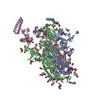
|
|---|---|
| 1 |
|
-Components
| #1: Protein | Mass: 142558.094 Da / Num. of mol.: 3 Source method: isolated from a genetically manipulated source Source: (gene. exp.) Severe acute respiratory syndrome coronavirus 2, (gene. exp.) Tequatrovirus T4Gene: S, 2 / Production host:  Homo sapiens (human) / References: UniProt: P0DTC2, UniProt: M1E1E4 Homo sapiens (human) / References: UniProt: P0DTC2, UniProt: M1E1E4#2: Protein | Mass: 8829.904 Da / Num. of mol.: 2 Source method: isolated from a genetically manipulated source Source: (gene. exp.) Drosophila melanogaster (fruit fly) / Gene: l(2)gd1, lgd, CG4713 / Production host:  Escherichia coli (E. coli) Escherichia coli (E. coli)#3: Polysaccharide | 2-acetamido-2-deoxy-beta-D-glucopyranose-(1-4)-2-acetamido-2-deoxy-beta-D-glucopyranose / Mass: 424.401 Da / Num. of mol.: 18Source method: isolated from a genetically manipulated source #4: Polysaccharide | beta-D-mannopyranose-(1-4)-2-acetamido-2-deoxy-beta-D-glucopyranose-(1-4)-2-acetamido-2-deoxy-beta- ...beta-D-mannopyranose-(1-4)-2-acetamido-2-deoxy-beta-D-glucopyranose-(1-4)-2-acetamido-2-deoxy-beta-D-glucopyranose / Mass: 586.542 Da / Num. of mol.: 12Source method: isolated from a genetically manipulated source #5: Sugar | ChemComp-NAG / N-Acetylglucosamine  Type: D-saccharide, beta linking / Mass: 221.208 Da / Num. of mol.: 16 / Source method: obtained synthetically / Formula: C8H15NO6 Type: D-saccharide, beta linking / Mass: 221.208 Da / Num. of mol.: 16 / Source method: obtained synthetically / Formula: C8H15NO6Has ligand of interest | N | |
|---|
-Experimental details
-Experiment
| Experiment | Method: ELECTRON MICROSCOPY |
|---|---|
| EM experiment | Aggregation state: PARTICLE / 3D reconstruction method: single particle reconstruction |
- Sample preparation
| Component |
| ||||||||||||||||||||||||
|---|---|---|---|---|---|---|---|---|---|---|---|---|---|---|---|---|---|---|---|---|---|---|---|---|---|
| Molecular weight |
| ||||||||||||||||||||||||
| Source (natural) |
| ||||||||||||||||||||||||
| Source (recombinant) |
| ||||||||||||||||||||||||
| Buffer solution | pH: 7.5 | ||||||||||||||||||||||||
| Specimen | Conc.: 1 mg/ml / Embedding applied: NO / Shadowing applied: NO / Staining applied: NO / Vitrification applied: YES | ||||||||||||||||||||||||
| Specimen support | Grid material: COPPER / Grid mesh size: 400 divisions/in. / Grid type: Quantifoil R2/1 | ||||||||||||||||||||||||
| Vitrification | Instrument: FEI VITROBOT MARK IV / Cryogen name: ETHANE / Humidity: 90 % / Chamber temperature: 298 K |
- Electron microscopy imaging
| Experimental equipment |  Model: Titan Krios / Image courtesy: FEI Company |
|---|---|
| Microscopy | Model: FEI TITAN KRIOS |
| Electron gun | Electron source: FIELD EMISSION GUN / Accelerating voltage: 300 kV / Illumination mode: FLOOD BEAM |
| Electron lens | Mode: BRIGHT FIELDBright-field microscopy / Nominal magnification: 165000 X / Nominal defocus max: 2000 nm / Nominal defocus min: 800 nm / Cs: 2.7 mm / Alignment procedure: COMA FREE |
| Specimen holder | Cryogen: NITROGEN / Specimen holder model: FEI TITAN KRIOS AUTOGRID HOLDER |
| Image recording | Average exposure time: 5.82 sec. / Electron dose: 60 e/Å2 / Film or detector model: FEI FALCON IV (4k x 4k) |
| EM imaging optics | Energyfilter name: TFS Selectris X |
- Processing
| Software | Name: PHENIX / Version: 1.19.2_4158: / Classification: refinement | ||||||||||||||||||||||||||||||
|---|---|---|---|---|---|---|---|---|---|---|---|---|---|---|---|---|---|---|---|---|---|---|---|---|---|---|---|---|---|---|---|
| EM software |
| ||||||||||||||||||||||||||||||
| CTF correction | Type: NONE | ||||||||||||||||||||||||||||||
| Particle selection | Num. of particles selected: 1820333 | ||||||||||||||||||||||||||||||
| Symmetry | Point symmetry: C1 (asymmetric) | ||||||||||||||||||||||||||||||
| 3D reconstruction | Resolution: 2.8 Å / Resolution method: FSC 0.143 CUT-OFF / Num. of particles: 50758 / Algorithm: BACK PROJECTION / Symmetry type: POINT | ||||||||||||||||||||||||||||||
| Atomic model building | B value: 33.8 / Protocol: AB INITIO MODEL / Space: REAL | ||||||||||||||||||||||||||||||
| Atomic model building | PDB-ID: 7QO7 Accession code: 7QO7 / Source name: PDB / Type: experimental model | ||||||||||||||||||||||||||||||
| Refine LS restraints |
|