 Movie
Movie Controller

[English] 日本語
 Yorodumi
- PDB-7zla: Cryo-EM structure of holo-PdxR from Bacillus clausii bound to its... -
+ Open data

- Basic information
| Entry | Database: PDB / ID: 7zla | ||||||
|---|---|---|---|---|---|---|---|
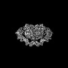
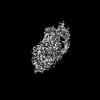
| Title | Cryo-EM structure of holo-PdxR from Bacillus clausii bound to its target DNA in the half-closed conformation | ||||||
 Components Components |
| ||||||
 Keywords Keywords |  DNA BINDING PROTEIN / transcription factor / PLP-binding protein / domain-swap homodimer DNA BINDING PROTEIN / transcription factor / PLP-binding protein / domain-swap homodimer | ||||||
| Function / homology |  Function and homology informationtransaminase activity / biosynthetic process / pyridoxal phosphate binding / DNA-binding transcription factor activity Function and homology informationtransaminase activity / biosynthetic process / pyridoxal phosphate binding / DNA-binding transcription factor activitySimilarity search - Function | ||||||
| Biological species |  Alkalihalobacillus clausii (bacteria) Alkalihalobacillus clausii (bacteria) | ||||||
| Method | ELECTRON MICROSCOPY / single particle reconstruction / cryo EM / Resolution: 3.99 Å | ||||||
 Authors Authors | Freda, I. / Montemiglio, L.C. / Tramonti, A. / Contestabile, R. / Vallone, B. / Savino, C. / Exertier, C. / Bolognesi, M. / Chaves Sanjuan, A. | ||||||
| Funding support | 1items
| ||||||
 Citation Citation | Journal: Nucleic Acids Res / Year: 2023 Title: Structural insights into the DNA recognition mechanism by the bacterial transcription factor PdxR. Authors: Ida Freda / Cécile Exertier / Anna Barile / Antonio Chaves-Sanjuan / Mirella Vivoli Vega / Michail N Isupov / Nicholas J Harmer / Elena Gugole / Paolo Swuec / Martino Bolognesi / Anita ...Authors: Ida Freda / Cécile Exertier / Anna Barile / Antonio Chaves-Sanjuan / Mirella Vivoli Vega / Michail N Isupov / Nicholas J Harmer / Elena Gugole / Paolo Swuec / Martino Bolognesi / Anita Scipioni / Carmelinda Savino / Martino Luigi Di Salvo / Roberto Contestabile / Beatrice Vallone / Angela Tramonti / Linda Celeste Montemiglio /   Abstract: Specificity in protein-DNA recognition arises from the synergy of several factors that stem from the structural and chemical signatures encoded within the targeted DNA molecule. Here, we deciphered ...Specificity in protein-DNA recognition arises from the synergy of several factors that stem from the structural and chemical signatures encoded within the targeted DNA molecule. Here, we deciphered the nature of the interactions driving DNA recognition and binding by the bacterial transcription factor PdxR, a member of the MocR family responsible for the regulation of pyridoxal 5'-phosphate (PLP) biosynthesis. Single particle cryo-EM performed on the PLP-PdxR bound to its target DNA enabled the isolation of three conformers of the complex, which may be considered as snapshots of the binding process. Moreover, the resolution of an apo-PdxR crystallographic structure provided a detailed description of the transition of the effector domain to the holo-PdxR form triggered by the binding of the PLP effector molecule. Binding analyses of mutated DNA sequences using both wild type and PdxR variants revealed a central role of electrostatic interactions and of the intrinsic asymmetric bending of the DNA in allosterically guiding the holo-PdxR-DNA recognition process, from the first encounter through the fully bound state. Our results detail the structure and dynamics of the PdxR-DNA complex, clarifying the mechanism governing the DNA-binding mode of the holo-PdxR and the regulation features of the MocR family of transcription factors. | ||||||
| History |
|
- Structure visualization
| Structure viewer | Molecule: MolmilJmol/JSmol |
|---|
- Downloads & links
-Download
| PDBx/mmCIF format | 7zla.cif.gz | 206.2 KB | Display | PDBx/mmCIF format |
|---|---|---|---|---|
| PDB format | pdb7zla.ent.gz | 157.5 KB | Display | PDB format |
| PDBx/mmJSON format | 7zla.json.gz | Tree view | PDBx/mmJSON format | |
| Others |  Other downloads Other downloads |
-Validation report
| Arichive directory | https://data.pdbj.org/pub/pdb/validation_reports/zl/7zlaftp://data.pdbj.org/pub/pdb/validation_reports/zl/7zla | HTTPS FTP |
|---|
-Related structure data
| Related structure data |  14778MC 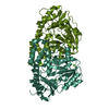 7pq9C 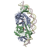 7zn5C 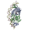 7zpaC 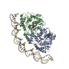 7zthC M: map data used to model this data C: citing same article ( |
|---|---|
| Similar structure data |

-Links
 PDBj
PDBj


- Assembly
| Deposited unit | 
|
|---|---|
| 1 |
|
-Components
| #1: Protein | Mass: 55499.121 Da / Num. of mol.: 2 Source method: isolated from a genetically manipulated source Source: (gene. exp.) Alkalihalobacillus clausii (bacteria) / Gene: CHH72_17460 / Production host: Escherichia coli BL21(DE3) (bacteria) / References: UniProt: A0A268NVG2#2: DNA chain | | Mass: 14667.447 Da / Num. of mol.: 1 / Source method: obtained synthetically / Source: (synth.) Alkalihalobacillus clausii (bacteria)#3: DNA chain | | Mass: 14894.607 Da / Num. of mol.: 1 / Source method: obtained synthetically / Source: (synth.) Alkalihalobacillus clausii (bacteria)Has ligand of interest | N | |
|---|
-Experimental details
-Experiment
| Experiment | Method: ELECTRON MICROSCOPY |
|---|---|
| EM experiment | Aggregation state: PARTICLE / 3D reconstruction method: single particle reconstruction |
- Sample preparation
| Component | Name: Binary complex between the PLP-bound homodimeric PdxR and a 48bp double strand DNA fragment Type: COMPLEX / Entity ID: all / Source: RECOMBINANT | ||||||||||||
|---|---|---|---|---|---|---|---|---|---|---|---|---|---|
| Molecular weight | Value: 0.136 MDa / Experimental value: NO | ||||||||||||
| Source (natural) | Organism: Alkalihalobacillus clausii (bacteria) | ||||||||||||
| Source (recombinant) | Organism: Escherichia coli BL21(DE3) (bacteria) | ||||||||||||
| Buffer solution | pH: 7.5 | ||||||||||||
| Buffer component |
| ||||||||||||
| Specimen | Conc.: 0.5 mg/ml / Embedding applied: NO / Shadowing applied: NO / Staining applied: NO / Vitrification applied: YES | ||||||||||||
| Specimen support | Grid material: COPPER / Grid mesh size: 300 divisions/in. / Grid type: Quantifoil R1.2/1.3 | ||||||||||||
| Vitrification | Instrument: FEI VITROBOT MARK IV / Cryogen name: ETHANE / Humidity: 100 % / Chamber temperature: 277.15 K |
- Electron microscopy imaging
| Experimental equipment |  Model: Talos Arctica / Image courtesy: FEI Company |
|---|---|
| Microscopy | Model: FEI TALOS ARCTICA |
| Electron gun | Electron source: FIELD EMISSION GUN / Accelerating voltage: 200 kV / Illumination mode: FLOOD BEAM |
| Electron lens | Mode: BRIGHT FIELDBright-field microscopy / Nominal magnification: 120000 X / Nominal defocus max: 2200 nm / Nominal defocus min: 800 nm |
| Specimen holder | Cryogen: NITROGEN |
| Image recording | Average exposure time: 1 sec. / Electron dose: 40 e/Å2 / Detector mode: COUNTING / Film or detector model: FEI FALCON III (4k x 4k) / Num. of grids imaged: 1 / Num. of real images: 3284 |
- Processing
| Software | Name: PHENIX / Version: 1.19.2_4158: / Classification: refinement | ||||||||||||||||||||||||||||||||||||
|---|---|---|---|---|---|---|---|---|---|---|---|---|---|---|---|---|---|---|---|---|---|---|---|---|---|---|---|---|---|---|---|---|---|---|---|---|---|
| EM software |
| ||||||||||||||||||||||||||||||||||||
| CTF correction | Type: PHASE FLIPPING ONLY | ||||||||||||||||||||||||||||||||||||
| Particle selection | Num. of particles selected: 1358491 | ||||||||||||||||||||||||||||||||||||
| Symmetry | Point symmetry: C1 (asymmetric) | ||||||||||||||||||||||||||||||||||||
| 3D reconstruction | Resolution: 3.99 Å / Resolution method: FSC 0.143 CUT-OFF / Num. of particles: 68689 / Symmetry type: POINT | ||||||||||||||||||||||||||||||||||||
| Atomic model building | B value: 181 / Protocol: RIGID BODY FIT / Space: REAL | ||||||||||||||||||||||||||||||||||||
| Atomic model building | PDB-ID: 7PQ9 Accession code: 7PQ9 / Source name: PDB / Type: experimental model | ||||||||||||||||||||||||||||||||||||
| Refinement | Cross valid method: NONE Stereochemistry target values: GeoStd + Monomer Library + CDL v1.2 | ||||||||||||||||||||||||||||||||||||
| Displacement parameters | Biso mean: 74.69 Å2 | ||||||||||||||||||||||||||||||||||||
| Refine LS restraints |
|
