 Movie
Movie Controller

[English] 日本語
 Yorodumi
- PDB-7zjz: catalytically non active S532A mutant of oligopeptidase B from S.... -
+ Open data

- Basic information
| Entry | Database: PDB / ID: 7zjz | ||||||
|---|---|---|---|---|---|---|---|
| Title | catalytically non active S532A mutant of oligopeptidase B from S. proteomaculans | ||||||
 Components Components | Oligopeptidase B | ||||||
 Keywords Keywords | HYDROLASE | ||||||
| Function / homology |  Function and homology information Function and homology information | ||||||
| Biological species |  Serratia proteamaculans (bacteria) Serratia proteamaculans (bacteria) | ||||||
| Method | X-RAY DIFFRACTION / SYNCHROTRON / MOLECULAR REPLACEMENT / Resolution: 1.9 Å | ||||||
 Authors Authors | Petrenko, D.E. / Boyko, K.M. / Nikolaeva, A.Y. / Vlaskina, A.V. / Mikhailova, A.G. / Timofeev, V.I. / Rakitina, T.V. | ||||||
| Funding support | 1items
| ||||||
 Citation Citation | Journal: Crystals / Year: 2022 Title: Elucidation of the Conformational Transition of Oligopeptidase B by an Integrative Approach Based on the Combination of X-ray, SAXS, and Essential Dynamics Sampling Simulation Authors: Britikov, V.V. / Timofeev, V.I. / Petrenko, D.E. / Britikova, E.V. / Nikolaeva, A.Y. / Vlaskina, A.V. / Boyko, K.M. / Mikhailova, A.G. / Rakitina, T.V. | ||||||
| History |
|
- Structure visualization
| Structure viewer | Molecule: MolmilJmol/JSmol |
|---|
- Downloads & links
-Download
| PDBx/mmCIF format | 7zjz.cif.gz | 160.9 KB | Display | PDBx/mmCIF format |
|---|---|---|---|---|
| PDB format | pdb7zjz.ent.gz | 124.2 KB | Display | PDB format |
| PDBx/mmJSON format | 7zjz.json.gz | Tree view | PDBx/mmJSON format | |
| Others |  Other downloads Other downloads |
-Validation report
| Arichive directory | https://data.pdbj.org/pub/pdb/validation_reports/zj/7zjzftp://data.pdbj.org/pub/pdb/validation_reports/zj/7zjz | HTTPS FTP |
|---|
-Related structure data
| Related structure data |  7ywsC 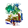 7yx7C  6tf5 S: Starting model for refinement C: citing same article ( |
|---|---|
| Similar structure data |
-Links
 PDBj
PDBj


- Assembly
| Deposited unit | 
| ||||||||
|---|---|---|---|---|---|---|---|---|---|
| 1 |
| ||||||||
| Unit cell |
|
-Components
| #1: Protein | / Prolyl oligopeptidase family serine peptidase Mass: 78382.797 Da / Num. of mol.: 1 Source method: isolated from a genetically manipulated source Source: (gene. exp.) Serratia proteamaculans (bacteria) / Gene: opdB, JKX24_00380 / Production host: Escherichia coli (E. coli) / References: UniProt: B3VI58 | ||||
|---|---|---|---|---|---|
| #2: Chemical | ChemComp-SPM / Spermine  Mass: 202.340 Da / Num. of mol.: 4 / Source method: obtained synthetically / Formula: C10H26N4 Mass: 202.340 Da / Num. of mol.: 4 / Source method: obtained synthetically / Formula: C10H26N4#3: Water | ChemComp-HOH / | Water Mass: 18.015 Da / Num. of mol.: 368 / Source method: isolated from a natural source / Formula: H2O Mass: 18.015 Da / Num. of mol.: 368 / Source method: isolated from a natural source / Formula: H2OHas ligand of interest | N | |
-Experimental details
-Experiment
| Experiment | Method: X-RAY DIFFRACTION / Number of used crystals: 1 |
|---|
- Sample preparation
| Crystal | Density Matthews: 2.54 Å3/Da / Density % sol: 51.62 % |
|---|---|
| Crystal grow | Temperature: 277 K / Method: counter-diffusion Details: 200 mM Lithium sulfate, 100 mM Bis-Tris pH 5.5, 23% PEG 3350 |
-Data collection
| Diffraction | Mean temperature: 100 K / Serial crystal experiment: N |
|---|---|
| Diffraction source | Source: SYNCHROTRON / Site: SPring-8  / Beamline: BL41XU / Wavelength: 0.7 Å / Beamline: BL41XU / Wavelength: 0.7 Å |
| Detector | Type: DECTRIS EIGER X 16M / Detector: PIXEL / Date: Oct 10, 2019 |
| Radiation | Protocol: SINGLE WAVELENGTH / Monochromatic (M) / Laue (L): M / Scattering type: x-ray |
| Radiation wavelength | Wavelength: 0.7 Å / Relative weight: 1 |
| Reflection | Resolution: 1.9→30 Å / Num. obs: 63693 / % possible obs: 99.96 % / Redundancy: 7.15 % / Rmerge(I) obs: 0.115 / Rrim(I) all: 0.124 / Net I/σ(I): 3.3271 |
| Reflection shell | Resolution: 1.9→2 Å / Redundancy: 2.27 % / Rmerge(I) obs: 0.57 / Mean I/σ(I) obs: 2.02 / Num. unique obs: 9183 / Rrim(I) all: 0.61 / % possible all: 100 |
- Processing
| Software |
| ||||||||||||||||||||||||||||||||||||||||||||||||||||||||||||
|---|---|---|---|---|---|---|---|---|---|---|---|---|---|---|---|---|---|---|---|---|---|---|---|---|---|---|---|---|---|---|---|---|---|---|---|---|---|---|---|---|---|---|---|---|---|---|---|---|---|---|---|---|---|---|---|---|---|---|---|---|---|
| Refinement | Method to determine structure: MOLECULAR REPLACEMENT Starting model: 6TF5 6tf5 Resolution: 1.9→29.44 Å / Cor.coef. Fo:Fc: 0.963 / Cor.coef. Fo:Fc free: 0.936 / SU B: 4.598 / SU ML: 0.127 / Cross valid method: THROUGHOUT / σ(F): 0 / ESU R: 0.154 / ESU R Free: 0.152 / Stereochemistry target values: MAXIMUM LIKELIHOOD Details: HYDROGENS HAVE BEEN ADDED IN THE RIDING POSITIONS U VALUES : REFINED INDIVIDUALLY
| ||||||||||||||||||||||||||||||||||||||||||||||||||||||||||||
| Solvent computation | Ion probe radii: 0.8 Å / Shrinkage radii: 0.8 Å / VDW probe radii: 1.2 Å / Solvent model: MASK | ||||||||||||||||||||||||||||||||||||||||||||||||||||||||||||
| Displacement parameters | Biso max: 131.06 Å2 / Biso mean: 40.059 Å2 / Biso min: 19.49 Å2
| ||||||||||||||||||||||||||||||||||||||||||||||||||||||||||||
| Refinement step | Cycle: final / Resolution: 1.9→29.44 Å
| ||||||||||||||||||||||||||||||||||||||||||||||||||||||||||||
| Refine LS restraints |
| ||||||||||||||||||||||||||||||||||||||||||||||||||||||||||||
| LS refinement shell | Resolution: 1.9→1.949 Å / Rfactor Rfree error: 0 / Total num. of bins used: 20
|
