 Movie
Movie Controller

[English] 日本語
 Yorodumi
- PDB-7zjf: R399E, a mutated form of GDF5, for disease modification of osteoa... -
+ Open data

- Basic information
| Entry | Database: PDB / ID: 7zjf | ||||||
|---|---|---|---|---|---|---|---|
| Title | R399E, a mutated form of GDF5, for disease modification of osteoarthritis | ||||||
 Components Components | Growth/differentiation factor 5 | ||||||
 Keywords Keywords |  SIGNALING PROTEIN / GROWTH FACTOR / GROWTH DIFFERENTIATION FACTOR / BONE MORPHOGENETIC SIGNALING PROTEIN / GROWTH FACTOR / GROWTH DIFFERENTIATION FACTOR / BONE MORPHOGENETIC | ||||||
| Function / homology |  Function and homology information Function and homology informationossification involved in bone remodeling / forelimb morphogenesis / BMP binding / chondroblast differentiation / hindlimb morphogenesis / negative regulation of mesenchymal cell apoptotic process / positive regulation of chondrocyte differentiation / mesenchymal cell apoptotic process / positive regulation of BMP signaling pathway / negative regulation of chondrocyte differentiation ...ossification involved in bone remodeling / forelimb morphogenesis / BMP binding / chondroblast differentiation / hindlimb morphogenesis / negative regulation of mesenchymal cell apoptotic process / positive regulation of chondrocyte differentiation / mesenchymal cell apoptotic process / positive regulation of BMP signaling pathway / negative regulation of chondrocyte differentiation / embryonic limb morphogenesis / Molecules associated with elastic fibres / positive regulation of SMAD protein signal transduction / regulation of multicellular organism growth / chondrocyte differentiation / BMP signaling pathway / response to mechanical stimulus / positive regulation of neuron differentiation / transforming growth factor beta receptor signaling pathway / cytokine activity / growth factor activity / negative regulation of epithelial cell proliferation / cell-cell signaling / negative regulation of neuron apoptotic process / extracellular space / extracellular region / identical protein binding / plasma membraneSimilarity search - Function | ||||||
| Biological species |  Homo sapiens (human) Homo sapiens (human) | ||||||
| Method | X-RAY DIFFRACTION / SYNCHROTRON / MOLECULAR REPLACEMENT / Resolution: 1.3 Å | ||||||
 Authors Authors | Gigout, A. / Wekmann, D. / Menges, S. / Brenneis, C. / Henson, F. / Cowan, K.J. / Musil, D. / Thudium, C. / Guehring, H. / Michaelis, M. / Kleinschmidt-Doerr, K. | ||||||
| Funding support | 1items
| ||||||
 Citation Citation | Journal: Arthritis Rheumatol / Year: 2023 Title: R399E, A Mutated Form of Growth and Differentiation Factor 5, for Disease Modification of Osteoarthritis. Authors: Gigout, A. / Werkmann, D. / Menges, S. / Brenneis, C. / Henson, F. / Cowan, K.J. / Musil, D. / Thudium, C.S. / Guhring, H. / Michaelis, M. / Kleinschmidt-Doerr, K. | ||||||
| History |
|
- Structure visualization
| Structure viewer | Molecule: MolmilJmol/JSmol |
|---|
- Downloads & links
-Download
| PDBx/mmCIF format | 7zjf.cif.gz | 109.2 KB | Display | PDBx/mmCIF format |
|---|---|---|---|---|
| PDB format | pdb7zjf.ent.gz | 82.9 KB | Display | PDB format |
| PDBx/mmJSON format | 7zjf.json.gz | Tree view | PDBx/mmJSON format | |
| Others |  Other downloads Other downloads |
-Validation report
| Arichive directory | https://data.pdbj.org/pub/pdb/validation_reports/zj/7zjfftp://data.pdbj.org/pub/pdb/validation_reports/zj/7zjf | HTTPS FTP |
|---|
-Related structure data
| Related structure data | 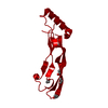 2bhkS S: Starting model for refinement |
|---|---|
| Similar structure data |
-Links
 PDBj
PDBj


- Assembly
| Deposited unit | 
| ||||||||
|---|---|---|---|---|---|---|---|---|---|
| 1 |
| ||||||||
| Unit cell |
|
-Components
| #1: Protein | Mass: 13570.558 Da / Num. of mol.: 2 Source method: isolated from a genetically manipulated source Source: (gene. exp.) Homo sapiens (human) / Gene: GDF5, BMP14, CDMP1 / Production host:  Escherichia coli (E. coli) / References: UniProt: P43026 Escherichia coli (E. coli) / References: UniProt: P43026#2: Water | ChemComp-HOH / | Water Mass: 18.015 Da / Num. of mol.: 306 / Source method: isolated from a natural source / Formula: H2O Mass: 18.015 Da / Num. of mol.: 306 / Source method: isolated from a natural source / Formula: H2O |
|---|
-Experimental details
-Experiment
| Experiment | Method: X-RAY DIFFRACTION / Number of used crystals: 1 |
|---|
- Sample preparation
| Crystal | Density Matthews: 2.8 Å3/Da / Density % sol: 56.05 % |
|---|---|
| Crystal grow | Temperature: 293 K / Method: vapor diffusion, hanging drop / pH: 8.15 / Details: 01 M Hepes, 32% PEG 400, 230 mM MgCl2, pH 8.15 |
-Data collection
| Diffraction | Mean temperature: 100 K / Serial crystal experiment: N |
|---|---|
| Diffraction source | Source: SYNCHROTRON / Site: SLS  / Beamline: X06SA / Wavelength: 1.00003 Å / Beamline: X06SA / Wavelength: 1.00003 Å |
| Detector | Type: DECTRIS PILATUS 2M-F / Detector: PIXEL / Date: Aug 26, 2015 |
| Radiation | Protocol: SINGLE WAVELENGTH / Monochromatic (M) / Laue (L): M / Scattering type: x-ray |
| Radiation wavelength | Wavelength: 1.00003 Å / Relative weight: 1 |
| Reflection | Resolution: 1.3→49.22 Å / Num. obs: 75754 / % possible obs: 99.9 % / Redundancy: 6.5 % / CC1/2: 0.999 / Rpim(I) all: 0.023 / Rrim(I) all: 0.069 / Net I/σ(I): 11.6 |
| Reflection shell | Resolution: 1.3→1.38 Å / Redundancy: 6.1 % / Num. unique obs: 11997 / CC1/2: 0.516 / Rpim(I) all: 0.436 / % possible all: 99.2 |
- Processing
| Software |
| ||||||||||||||||||||||||||||||||||||||||||||||||||||||||||||||||||||||||||||||||||||||||||||||||||||||||||||
|---|---|---|---|---|---|---|---|---|---|---|---|---|---|---|---|---|---|---|---|---|---|---|---|---|---|---|---|---|---|---|---|---|---|---|---|---|---|---|---|---|---|---|---|---|---|---|---|---|---|---|---|---|---|---|---|---|---|---|---|---|---|---|---|---|---|---|---|---|---|---|---|---|---|---|---|---|---|---|---|---|---|---|---|---|---|---|---|---|---|---|---|---|---|---|---|---|---|---|---|---|---|---|---|---|---|---|---|---|---|
| Refinement | Method to determine structure: MOLECULAR REPLACEMENT Starting model: 2bhk Resolution: 1.3→49.22 Å / Cor.coef. Fo:Fc: 0.952 / Cor.coef. Fo:Fc free: 0.949 / SU R Cruickshank DPI: 0.048 / Cross valid method: THROUGHOUT / σ(F): 0 / SU R Blow DPI: 0.052 / SU Rfree Blow DPI: 0.052 / SU Rfree Cruickshank DPI: 0.048
| ||||||||||||||||||||||||||||||||||||||||||||||||||||||||||||||||||||||||||||||||||||||||||||||||||||||||||||
| Displacement parameters | Biso max: 77.6 Å2 / Biso mean: 31.25 Å2 / Biso min: 17.19 Å2
| ||||||||||||||||||||||||||||||||||||||||||||||||||||||||||||||||||||||||||||||||||||||||||||||||||||||||||||
| Refine analyze | Luzzati coordinate error obs: 0.41 Å | ||||||||||||||||||||||||||||||||||||||||||||||||||||||||||||||||||||||||||||||||||||||||||||||||||||||||||||
| Refinement step | Cycle: final / Resolution: 1.3→49.22 Å
| ||||||||||||||||||||||||||||||||||||||||||||||||||||||||||||||||||||||||||||||||||||||||||||||||||||||||||||
| Refine LS restraints |
| ||||||||||||||||||||||||||||||||||||||||||||||||||||||||||||||||||||||||||||||||||||||||||||||||||||||||||||
| LS refinement shell | Resolution: 1.3→1.31 Å / Rfactor Rfree error: 0 / Total num. of bins used: 51
| ||||||||||||||||||||||||||||||||||||||||||||||||||||||||||||||||||||||||||||||||||||||||||||||||||||||||||||
| Refinement TLS params. | Method: refined / Refine-ID: X-RAY DIFFRACTION
| ||||||||||||||||||||||||||||||||||||||||||||||||||||||||||||||||||||||||||||||||||||||||||||||||||||||||||||
| Refinement TLS group |
|
