 Movie
Movie Controller

[English] 日本語
 Yorodumi
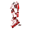
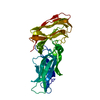
- PDB-2bhk: Crystal structure of human growth and differentiation factor 5 (GDF5) -
+ Open data

- Basic information
| Entry | Database: PDB / ID: 2bhk | ||||||
|---|---|---|---|---|---|---|---|
| Title | Crystal structure of human growth and differentiation factor 5 (GDF5) | ||||||
 Components Components | GROWTH DIFFERENTIATION FACTOR 5 | ||||||
 Keywords Keywords | GROWTH FACTOR / GROWTH DIFFERENTIATION FACTOR / BONE MORPHOGENETIC FACTOR / HORMONE-RECEPTOR INTERACTION / CYSTINE KNOT / PREFORMED RECEPTOR DIMER / CYTOKINE / DWARFISM / GLYCOPROTEIN | ||||||
| Function / homology |  Function and homology information Function and homology informationossification involved in bone remodeling / forelimb morphogenesis / BMP binding / chondroblast differentiation / hindlimb morphogenesis / negative regulation of mesenchymal cell apoptotic process / positive regulation of chondrocyte differentiation / mesenchymal cell apoptotic process / positive regulation of BMP signaling pathway / negative regulation of chondrocyte differentiation ...ossification involved in bone remodeling / forelimb morphogenesis / BMP binding / chondroblast differentiation / hindlimb morphogenesis / negative regulation of mesenchymal cell apoptotic process / positive regulation of chondrocyte differentiation / mesenchymal cell apoptotic process / positive regulation of BMP signaling pathway / negative regulation of chondrocyte differentiation / embryonic limb morphogenesis / Molecules associated with elastic fibres / positive regulation of SMAD protein signal transduction / regulation of multicellular organism growth / chondrocyte differentiation / BMP signaling pathway / response to mechanical stimulus / positive regulation of neuron differentiation / transforming growth factor beta receptor signaling pathway / cytokine activity / growth factor activity / negative regulation of epithelial cell proliferation / cell-cell signaling / negative regulation of neuron apoptotic process / extracellular space / extracellular region / identical protein binding / plasma membraneSimilarity search - Function | ||||||
| Biological species |  HOMO SAPIENS (human) HOMO SAPIENS (human) | ||||||
| Method | X-RAY DIFFRACTION / MOLECULAR REPLACEMENT / Resolution: 2.4 Å | ||||||
 Authors Authors | Schreuder, H. / Liesum, A. / Pohl, J. / Kruse, M. / Koyama, M. | ||||||
 Citation Citation | Journal: Biochem.Biophys.Res.Commun. / Year: 2005 Title: Crystal Structure of Recombinant Human Growth Differentiation Factor 5. Evidence for Interaction of the Type I and Type II Receptor Binding Sites. Authors: Schreuder, H. / Liesum, A. / Pohl, J. / Kruse, M. / Koyama, M. | ||||||
| History |
|
- Structure visualization
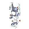
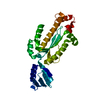
| Structure viewer | Molecule: MolmilJmol/JSmol |
|---|
- Downloads & links
-Download
| PDBx/mmCIF format | 2bhk.cif.gz | 36.9 KB | Display | PDBx/mmCIF format |
|---|---|---|---|---|
| PDB format | pdb2bhk.ent.gz | 24.3 KB | Display | PDB format |
| PDBx/mmJSON format | 2bhk.json.gz | Tree view | PDBx/mmJSON format | |
| Others |  Other downloads Other downloads |
-Validation report
| Arichive directory | https://data.pdbj.org/pub/pdb/validation_reports/bh/2bhkftp://data.pdbj.org/pub/pdb/validation_reports/bh/2bhk | HTTPS FTP |
|---|
-Related structure data
| Related structure data |  1bmpS S: Starting model for refinement |
|---|---|
| Similar structure data |


-Links
 PDBj
PDBj


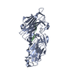
- Assembly
| Deposited unit | 
| ||||||||
|---|---|---|---|---|---|---|---|---|---|
| 1 | 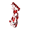
| ||||||||
| Unit cell |
|
-Components
| #1: Protein | / GDF-5 / CARTILAGE-DERIVED MORPHOGENETIC PROTEIN 1 / CDMP-1 Mass: 13598.638 Da / Num. of mol.: 1 Source method: isolated from a genetically manipulated source Details: INTERCHAIN DISULFIDE LINK BETWEEN TWO CRYSTALLOGRAPHICALLY RELATED MONOMERS Source: (gene. exp.) HOMO SAPIENS (human) / Plasmid: PUC18 / Production host:  ESCHERICHIA COLI (E. coli) / References: UniProt: P43026 ESCHERICHIA COLI (E. coli) / References: UniProt: P43026 | ||||
|---|---|---|---|---|---|
| #2: Chemical | ChemComp-IPA / Isopropyl alcohol  Mass: 60.095 Da / Num. of mol.: 5 / Source method: obtained synthetically / Formula: C3H8O / Comment: alkaloid*YM Mass: 60.095 Da / Num. of mol.: 5 / Source method: obtained synthetically / Formula: C3H8O / Comment: alkaloid*YM#3: Water | ChemComp-HOH / | Water Mass: 18.015 Da / Num. of mol.: 72 / Source method: isolated from a natural source / Formula: H2O Mass: 18.015 Da / Num. of mol.: 72 / Source method: isolated from a natural source / Formula: H2OCompound details | FUNCTION: POSSIBLE INVOLVEMEN | |
-Experimental details
-Experiment
| Experiment | Method: X-RAY DIFFRACTION / Number of used crystals: 1 |
|---|
- Sample preparation
| Crystal | Density Matthews: 5.1 Å3/Da / Density % sol: 75 % |
|---|---|
| Crystal grow | Method: vapor diffusion, hanging drop / pH: 5 Details: 100 MM SODIUM ACETATE, PH 5.0 30% ISOPROPANOL 14 MG/ML PROTEIN HANGING DROP |
-Data collection
| Diffraction | Mean temperature: 293 K |
|---|---|
| Diffraction source | Source: ROTATING ANODE / Type: ENRAF-NONIUS FR571 / Wavelength: 1.5418 |
| Detector | Type: MAR scanner 345 mm plate / Detector: IMAGE PLATE / Date: Aug 26, 1997 / Details: MIRRORS |
| Radiation | Monochromator: OSMIC MIRRORS / Protocol: SINGLE WAVELENGTH / Monochromatic (M) / Laue (L): M / Scattering type: x-ray |
| Radiation wavelength | Wavelength: 1.5418 Å / Relative weight: 1 |
| Reflection | Resolution: 2.4→60 Å / Num. obs: 9310 / % possible obs: 95.7 % / Redundancy: 3.7 % / Biso Wilson estimate: 9.2 Å2 / Rmerge(I) obs: 0.07 / Net I/σ(I): 17 |
| Reflection shell | Resolution: 2.4→2.5 Å / Redundancy: 3.6 % / Rmerge(I) obs: 0.28 / Mean I/σ(I) obs: 4.8 / % possible all: 98.6 |
- Processing
| Software |
| ||||||||||||||||||||||||||||||||||||||||||||||||||||||||||||
|---|---|---|---|---|---|---|---|---|---|---|---|---|---|---|---|---|---|---|---|---|---|---|---|---|---|---|---|---|---|---|---|---|---|---|---|---|---|---|---|---|---|---|---|---|---|---|---|---|---|---|---|---|---|---|---|---|---|---|---|---|---|
| Refinement | Method to determine structure: MOLECULAR REPLACEMENT Starting model: PDB ENTRY 1BMP Resolution: 2.4→8 Å / Data cutoff high absF: 1000000000 / Data cutoff low absF: 0.1 / Cross valid method: THROUGHOUT
| ||||||||||||||||||||||||||||||||||||||||||||||||||||||||||||
| Displacement parameters | Biso mean: 35.6 Å2 | ||||||||||||||||||||||||||||||||||||||||||||||||||||||||||||
| Refinement step | Cycle: LAST / Resolution: 2.4→8 Å
| ||||||||||||||||||||||||||||||||||||||||||||||||||||||||||||
| Refine LS restraints |
| ||||||||||||||||||||||||||||||||||||||||||||||||||||||||||||
| LS refinement shell | Resolution: 2.4→2.51 Å / Total num. of bins used: 8 /
|
