 Movie
Movie Controller


+ Open data

- Basic information
| Entry | Database: PDB / ID: 7yzv | ||||||
|---|---|---|---|---|---|---|---|
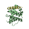
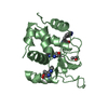
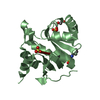
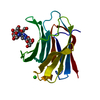
| Title | Ryegrass mottle virus serine protease domain S159A mutant | ||||||
 Components Components | RNA-directed RNA polymerase RNA-dependent RNA polymerase RNA-dependent RNA polymerase | ||||||
 Keywords Keywords | VIRAL PROTEIN / serine protease / viral protease / viral polypeptide / viral genome-linked protein | ||||||
| Function / homology |  Function and homology information Function and homology informationhost cell membrane / viral RNA genome replication / nucleotide binding / RNA-dependent RNA polymerase activity / serine-type endopeptidase activity / DNA-templated transcription / proteolysis / RNA binding / membraneSimilarity search - Function | ||||||
| Biological species |  Ryegrass mottle virus Ryegrass mottle virus | ||||||
| Method | X-RAY DIFFRACTION / SYNCHROTRON / MOLECULAR REPLACEMENT / Resolution: 1.6 Å | ||||||
 Authors Authors | Kalnins, G. | ||||||
| Funding support | European Union, 1items
| ||||||
 Citation Citation | Journal: Int J Mol Sci / Year: 2023 Title: VPg Impact on Ryegrass Mottle Virus Serine-like 3C Protease Proteolysis and Structure. Authors: Kalnins, G. / Ludviga, R. / Kalnciema, I. / Resevica, G. / Zeltina, V. / Bogans, J. / Tars, K. / Zeltins, A. / Balke, I. | ||||||
| History |
|
- Structure visualization
| Structure viewer | Molecule: MolmilJmol/JSmol |
|---|
- Downloads & links
-Download
| PDBx/mmCIF format | 7yzv.cif.gz | 59.5 KB | Display | PDBx/mmCIF format |
|---|---|---|---|---|
| PDB format | pdb7yzv.ent.gz | 33.8 KB | Display | PDB format |
| PDBx/mmJSON format | 7yzv.json.gz | Tree view | PDBx/mmJSON format | |
| Others |  Other downloads Other downloads |
-Validation report
| Arichive directory | https://data.pdbj.org/pub/pdb/validation_reports/yz/7yzvftp://data.pdbj.org/pub/pdb/validation_reports/yz/7yzv | HTTPS FTP |
|---|
-Related structure data
| Related structure data | 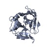 6fezC 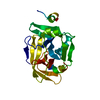 6ff0SC S: Starting model for refinement C: citing same article ( |
|---|---|
| Similar structure data |


-Links
 PDBj
PDBj- Assembly
| Deposited unit | 
| ||||||||||||
|---|---|---|---|---|---|---|---|---|---|---|---|---|---|
| 1 |
| ||||||||||||
| Unit cell |
| ||||||||||||
| Components on special symmetry positions |
|
-Components
| #1: Protein | RNA-dependent RNA polymerase Mass: 20867.982 Da / Num. of mol.: 1 / Mutation: S159A Source method: isolated from a genetically manipulated source Source: (gene. exp.) Ryegrass mottle virus / Production host:  Escherichia coli BL21(DE3) (bacteria) / References: UniProt: A0MCW0, RNA-directed RNA polymerase Escherichia coli BL21(DE3) (bacteria) / References: UniProt: A0MCW0, RNA-directed RNA polymerase |
|---|---|
| #2: Water | ChemComp-HOH / Water Mass: 18.015 Da / Num. of mol.: 99 / Source method: isolated from a natural source / Formula: H2O Mass: 18.015 Da / Num. of mol.: 99 / Source method: isolated from a natural source / Formula: H2O |
-Experimental details
-Experiment
| Experiment | Method: X-RAY DIFFRACTION / Number of used crystals: 1 |
|---|
- Sample preparation
| Crystal | Density Matthews: 2.78 Å3/Da / Density % sol: 55.85 % / Description: Needle-like |
|---|---|
| Crystal grow | Temperature: 293 K / Method: vapor diffusion, sitting drop / pH: 6.5 Details: 0.18 M ammonium sulfate, 22% PEG 8000, 0.1 M sodium cacodylate (pH 6.5) |
-Data collection
| Diffraction | Mean temperature: 90 K / Serial crystal experiment: N |
|---|---|
| Diffraction source | Source: SYNCHROTRON / Site: BESSY  / Beamline: 14.1 / Wavelength: 0.9184 Å / Beamline: 14.1 / Wavelength: 0.9184 Å |
| Detector | Type: DECTRIS PILATUS 6M / Detector: PIXEL / Date: Mar 10, 2020 |
| Radiation | Protocol: SINGLE WAVELENGTH / Monochromatic (M) / Laue (L): M / Scattering type: x-ray |
| Radiation wavelength | Wavelength: 0.9184 Å / Relative weight: 1 |
| Reflection | Resolution: 1.6→56.02 Å / Num. obs: 29492 / % possible obs: 95.9 % / Redundancy: 5.7 % / Biso Wilson estimate: 15.04 Å2 / CC1/2: 0.986 / Rmerge(I) obs: 0.148 / Net I/σ(I): 6.7 |
| Reflection shell | Resolution: 1.6→1.68 Å / Redundancy: 5.8 % / Rmerge(I) obs: 0.66 / Mean I/σ(I) obs: 2.2 / Num. unique obs: 4334 / CC1/2: 0.724 / % possible all: 97 |
- Processing
| Software |
| |||||||||||||||||||||||||||||||||||||||||||||||||||||||||||||||||||||||||||||
|---|---|---|---|---|---|---|---|---|---|---|---|---|---|---|---|---|---|---|---|---|---|---|---|---|---|---|---|---|---|---|---|---|---|---|---|---|---|---|---|---|---|---|---|---|---|---|---|---|---|---|---|---|---|---|---|---|---|---|---|---|---|---|---|---|---|---|---|---|---|---|---|---|---|---|---|---|---|---|
| Refinement | Method to determine structure: MOLECULAR REPLACEMENT Starting model: 6FF0 Resolution: 1.6→56.02 Å / SU ML: 0.2133 / Cross valid method: FREE R-VALUE / σ(F): 1.34 / Phase error: 26.4834 Stereochemistry target values: GeoStd + Monomer Library + CDL v1.2
| |||||||||||||||||||||||||||||||||||||||||||||||||||||||||||||||||||||||||||||
| Solvent computation | Shrinkage radii: 0.9 Å / VDW probe radii: 1.11 Å / Solvent model: FLAT BULK SOLVENT MODEL | |||||||||||||||||||||||||||||||||||||||||||||||||||||||||||||||||||||||||||||
| Displacement parameters | Biso mean: 18.13 Å2 | |||||||||||||||||||||||||||||||||||||||||||||||||||||||||||||||||||||||||||||
| Refinement step | Cycle: LAST / Resolution: 1.6→56.02 Å
| |||||||||||||||||||||||||||||||||||||||||||||||||||||||||||||||||||||||||||||
| Refine LS restraints |
| |||||||||||||||||||||||||||||||||||||||||||||||||||||||||||||||||||||||||||||
| LS refinement shell |
|
