 Movie
Movie Controller

[English] 日本語
 Yorodumi
- PDB-7yx3: Structure of the Mimivirus genomic fibre in its compact 6-start h... -
+ Open data

- Basic information
| Entry | Database: PDB / ID: 7yx3 | ||||||||||||||||||||||||
|---|---|---|---|---|---|---|---|---|---|---|---|---|---|---|---|---|---|---|---|---|---|---|---|---|---|
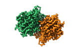
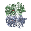
| Title | Structure of the Mimivirus genomic fibre in its compact 6-start helix form | ||||||||||||||||||||||||
 Components Components | Putative GMC-type oxidoreductase | ||||||||||||||||||||||||
 Keywords Keywords |  VIRAL PROTEIN / Mimivirus / Genomic fibre / Cytoplasmic infectious cycle / 1.2 Mb dsDNA / VIRUS VIRAL PROTEIN / Mimivirus / Genomic fibre / Cytoplasmic infectious cycle / 1.2 Mb dsDNA / VIRUS | ||||||||||||||||||||||||
| Function / homology |  Function and homology information Function and homology informationoxidoreductase activity, acting on CH-OH group of donors / flavin adenine dinucleotide bindingSimilarity search - Function | ||||||||||||||||||||||||
| Biological species |   Acanthamoeba polyphaga mimivirus Acanthamoeba polyphaga mimivirus | ||||||||||||||||||||||||
| Method | ELECTRON MICROSCOPY / helical reconstruction / cryo EM / Resolution: 4 Å | ||||||||||||||||||||||||
 Authors Authors | Villalta, A. / Schmitt, A. / Estrozi, L.F. / Quemin, E.R.J. / Alempic, J.M. / Lartigue, A. / Prazak, V. / Belmudes, L. / Vasishtan, D. / Colmant, A.M.G. ...Villalta, A. / Schmitt, A. / Estrozi, L.F. / Quemin, E.R.J. / Alempic, J.M. / Lartigue, A. / Prazak, V. / Belmudes, L. / Vasishtan, D. / Colmant, A.M.G. / Honore, F.A. / Coute, Y. / Grunewald, K. / Abergel, C. | ||||||||||||||||||||||||
| Funding support | European Union,  France, France,  Germany, Germany,  United Kingdom, 7items United Kingdom, 7items
| ||||||||||||||||||||||||
 Citation Citation | Journal: Elife / Year: 2022 Title: The giant mimivirus 1.2 Mb genome is elegantly organized into a 30-nm diameter helical protein shield. Authors: Alejandro Villalta / Alain Schmitt / Leandro F Estrozi / Emmanuelle R J Quemin / Jean-Marie Alempic / Audrey Lartigue / Vojtěch Pražák / Lucid Belmudes / Daven Vasishtan / Agathe M G ...Authors: Alejandro Villalta / Alain Schmitt / Leandro F Estrozi / Emmanuelle R J Quemin / Jean-Marie Alempic / Audrey Lartigue / Vojtěch Pražák / Lucid Belmudes / Daven Vasishtan / Agathe M G Colmant / Flora A Honoré / Yohann Couté / Kay Grünewald / Chantal Abergel / Abstract: Mimivirus is the prototype of the family of giant dsDNA viruses. Little is known about the organization of the 1.2 Mb genome inside the membrane-limited nucleoid filling the ~0.5 µm icosahedral ...Mimivirus is the prototype of the family of giant dsDNA viruses. Little is known about the organization of the 1.2 Mb genome inside the membrane-limited nucleoid filling the ~0.5 µm icosahedral capsids. Cryo-electron microscopy, cryo-electron tomography, and proteomics revealed that it is encased into a ~30-nm diameter helical protein shell surprisingly composed of two GMC-type oxidoreductases, which also form the glycosylated fibrils decorating the capsid. The genome is arranged in 5- or 6-start left-handed super-helices, with each DNA-strand lining the central channel. This luminal channel of the nucleoprotein fiber is wide enough to accommodate oxidative stress proteins and RNA polymerase subunits identified by proteomics. Such elegant supramolecular organization would represent a remarkable evolutionary strategy for packaging and protecting the genome, in a state ready for immediate transcription upon unwinding in the host cytoplasm. The parsimonious use of the same protein in two unrelated substructures of the virion is unexpected for a giant virus with thousand genes at its disposal. | ||||||||||||||||||||||||
| History |
|
- Structure visualization
| Structure viewer | Molecule: MolmilJmol/JSmol |
|---|
- Downloads & links
-Download
| PDBx/mmCIF format | 7yx3.cif.gz | 226.2 KB | Display | PDBx/mmCIF format |
|---|---|---|---|---|
| PDB format | pdb7yx3.ent.gz | 186.3 KB | Display | PDB format |
| PDBx/mmJSON format | 7yx3.json.gz | Tree view | PDBx/mmJSON format | |
| Others |  Other downloads Other downloads |
-Validation report
| Arichive directory | https://data.pdbj.org/pub/pdb/validation_reports/yx/7yx3ftp://data.pdbj.org/pub/pdb/validation_reports/yx/7yx3 | HTTPS FTP |
|---|
-Related structure data
| Related structure data |  14353MC 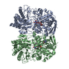 7ptvC 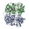 7yx4C 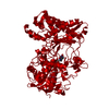 7yx5C C: citing same article ( M: map data used to model this data |
|---|---|
| Similar structure data |


-Links
 PDBj
PDBj
- Assembly
| Deposited unit | 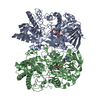
|
|---|---|
| 1 | x 9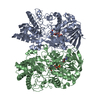
|
-Components
| #1: Protein | Mass: 77018.023 Da / Num. of mol.: 2 / Source method: isolated from a natural source / Source: (natural) Acanthamoeba polyphaga mimivirus / References: UniProt: A0A8A5IZP6#2: Chemical | Flavin adenine dinucleotide  Mass: 785.550 Da / Num. of mol.: 2 / Source method: obtained synthetically / Formula: C27H33N9O15P2 / Feature type: SUBJECT OF INVESTIGATION / Comment: FAD*YM Mass: 785.550 Da / Num. of mol.: 2 / Source method: obtained synthetically / Formula: C27H33N9O15P2 / Feature type: SUBJECT OF INVESTIGATION / Comment: FAD*YMHas ligand of interest | Y | |
|---|
-Experimental details
-Experiment
| Experiment | Method: ELECTRON MICROSCOPY |
|---|---|
| EM experiment | Aggregation state: FILAMENT / 3D reconstruction method: helical reconstruction |
- Sample preparation
| Component | Name: Mimivirus genomic fibre in its compact 6-start helix form Type: COMPLEX / Entity ID: #1 / Source: NATURAL |
|---|---|
| Molecular weight | Experimental value: NO |
| Source (natural) | Organism: Acanthamoeba polyphaga mimivirus / Strain: Reunion |
| Buffer solution | pH: 7.5 |
| Buffer component | Conc.: 40 mM / Name: Tris buffer / Formula: (HOCH2)3CNH2 |
| Specimen | Embedding applied: NO / Shadowing applied: NO / Staining applied: NO / Vitrification applied: YES |
| Vitrification | Cryogen name: ETHANE |
- Electron microscopy imaging
| Experimental equipment |  Model: Titan Krios / Image courtesy: FEI Company |
|---|---|
| Microscopy | Model: FEI TITAN KRIOS |
| Electron gun | Electron source: FIELD EMISSION GUN / Accelerating voltage: 300 kV / Illumination mode: OTHER |
| Electron lens | Mode: BRIGHT FIELDBright-field microscopy / Nominal defocus max: 3000 nm / Nominal defocus min: 1000 nm |
| Image recording | Electron dose: 50.6 e/Å2 / Film or detector model: GATAN K2 QUANTUM (4k x 4k) |
- Processing
| CTF correction | Type: PHASE FLIPPING AND AMPLITUDE CORRECTION | ||||||||||||||||||||||||
|---|---|---|---|---|---|---|---|---|---|---|---|---|---|---|---|---|---|---|---|---|---|---|---|---|---|
| Helical symmerty | Angular rotation/subunit: 49.43 ° / Axial rise/subunit: 20.47 Å / Axial symmetry: C3 | ||||||||||||||||||||||||
| 3D reconstruction | Resolution: 4 Å / Resolution method: FSC 0.5 CUT-OFF / Num. of particles: 8479 / Symmetry type: HELICAL | ||||||||||||||||||||||||
| Refine LS restraints |
|
