 Movie
Movie Controller

[English] 日本語
 Yorodumi
- PDB-7ycq: Crystal structure of human transthyretin variant A97S complexed w... -
+ Open data

- Basic information
| Entry | Database: PDB / ID: 7ycq | ||||||
|---|---|---|---|---|---|---|---|
| Title | Crystal structure of human transthyretin variant A97S complexed with Diflunisal | ||||||
 Components Components | Transthyretin | ||||||
 Keywords Keywords | TRANSPORT PROTEIN / Transport thyroxine | ||||||
| Function / homology |  Function and homology information Function and homology informationRetinoid cycle disease events / thyroid hormone binding / The canonical retinoid cycle in rods (twilight vision) / Non-integrin membrane-ECM interactions / purine nucleobase metabolic process / Retinoid metabolism and transport / hormone activity / azurophil granule lumen / Amyloid fiber formation / Neutrophil degranulation ...Retinoid cycle disease events / thyroid hormone binding / The canonical retinoid cycle in rods (twilight vision) / Non-integrin membrane-ECM interactions / purine nucleobase metabolic process / Retinoid metabolism and transport / hormone activity / azurophil granule lumen / Amyloid fiber formation / Neutrophil degranulation / extracellular space / extracellular exosome / extracellular region / identical protein bindingSimilarity search - Function | ||||||
| Biological species |  Homo sapiens (human) Homo sapiens (human) | ||||||
| Method | X-RAY DIFFRACTION / SYNCHROTRON / MOLECULAR REPLACEMENT / Resolution: 1.99 Å | ||||||
 Authors Authors | Wang, Y.S. / Huang, C.H. / Tzeng, S.R. | ||||||
| Funding support | 1items
| ||||||
 Citation Citation | Journal: Protein Sci. / Year: 2023 Title: A molecular basis for tetramer destabilization and aggregation of transthyretin Ala97Ser. Authors: Wang, Y.S. / Huang, C.H. / Liou, G.G. / Hsueh, H.W. / Liang, C.T. / Tseng, H.C. / Huang, S.J. / Chao, C.C. / Hsieh, S.T. / Tzeng, S.R. | ||||||
| History |
|
- Structure visualization
| Structure viewer | Molecule: MolmilJmol/JSmol |
|---|
- Downloads & links
-Download
| PDBx/mmCIF format | 7ycq.cif.gz | 107.6 KB | Display | PDBx/mmCIF format |
|---|---|---|---|---|
| PDB format | pdb7ycq.ent.gz | 80.5 KB | Display | PDB format |
| PDBx/mmJSON format | 7ycq.json.gz | Tree view | PDBx/mmJSON format | |
| Others |  Other downloads Other downloads |
-Validation report
| Arichive directory | https://data.pdbj.org/pub/pdb/validation_reports/yc/7ycqftp://data.pdbj.org/pub/pdb/validation_reports/yc/7ycq | HTTPS FTP |
|---|
-Related structure data
| Related structure data |  7y6jC 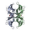 7ybrC  8hy4C  2qgbS S: Starting model for refinement C: citing same article ( |
|---|---|
| Similar structure data |
-Links
 PDBj
PDBj


- Assembly
| Deposited unit | 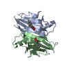
| ||||||||||||
|---|---|---|---|---|---|---|---|---|---|---|---|---|---|
| 1 | 
| ||||||||||||
| Unit cell |
|
-Components
| #1: Protein | / ATTR / Prealbumin / TBPA Mass: 13793.360 Da / Num. of mol.: 2 / Mutation: A97S Source method: isolated from a genetically manipulated source Source: (gene. exp.) Homo sapiens (human) / Gene: TTR, PALB / Production host:  Escherichia coli (E. coli) / References: UniProt: P02766 Escherichia coli (E. coli) / References: UniProt: P02766#2: Chemical | Diflunisal  Mass: 250.198 Da / Num. of mol.: 2 / Source method: obtained synthetically / Formula: C13H8F2O3 / Feature type: SUBJECT OF INVESTIGATION / Comment: antiinflammatory*YM Mass: 250.198 Da / Num. of mol.: 2 / Source method: obtained synthetically / Formula: C13H8F2O3 / Feature type: SUBJECT OF INVESTIGATION / Comment: antiinflammatory*YM#3: Water | ChemComp-HOH / | Water Mass: 18.015 Da / Num. of mol.: 97 / Source method: isolated from a natural source / Formula: H2O Mass: 18.015 Da / Num. of mol.: 97 / Source method: isolated from a natural source / Formula: H2OHas ligand of interest | Y | |
|---|
-Experimental details
-Experiment
| Experiment | Method: X-RAY DIFFRACTION / Number of used crystals: 1 |
|---|
- Sample preparation
| Crystal | Density Matthews: 2.11 Å3/Da / Density % sol: 41.75 % |
|---|---|
| Crystal grow | Temperature: 277 K / Method: vapor diffusion, hanging drop / pH: 5.5 Details: 1.5 M Ammonium Sulfate, 0.01M citrate buffer; soaked with 800 micromolar Dilfunisal |
-Data collection
| Diffraction | Mean temperature: 100 K / Serial crystal experiment: N |
|---|---|
| Diffraction source | Source: SYNCHROTRON / Site: NSRRC  / Beamline: BL13B1 / Wavelength: 1 Å / Beamline: BL13B1 / Wavelength: 1 Å |
| Detector | Type: ADSC QUANTUM 315r / Detector: CCD / Date: Nov 18, 2017 |
| Radiation | Protocol: SINGLE WAVELENGTH / Monochromatic (M) / Laue (L): M / Scattering type: x-ray |
| Radiation wavelength | Wavelength: 1 Å / Relative weight: 1 |
| Reflection | Resolution: 1.989→30 Å / Num. obs: 16347 / % possible obs: 99 % / Redundancy: 6.2 % / Biso Wilson estimate: 34.8 Å2 / Rmerge(I) obs: 0.055 / Rpim(I) all: 0.024 / Rrim(I) all: 0.061 / Net I/σ(I): 28.268292682927 |
| Reflection shell | Resolution: 1.99→2.06 Å / Rmerge(I) obs: 0.398 / Mean I/σ(I) obs: 3.5081967213115 / Num. unique obs: 1581 / CC1/2: 0.941 / CC star: 0.985 / Rpim(I) all: 0.195 / Rrim(I) all: 0.445 / % possible all: 98 |
- Processing
| Software |
| |||||||||||||||||||||||||||||||||||||||||||||||||
|---|---|---|---|---|---|---|---|---|---|---|---|---|---|---|---|---|---|---|---|---|---|---|---|---|---|---|---|---|---|---|---|---|---|---|---|---|---|---|---|---|---|---|---|---|---|---|---|---|---|---|
| Refinement | Method to determine structure: MOLECULAR REPLACEMENT Starting model: 2QGB Resolution: 1.99→22.26 Å / SU ML: 0.1895 / Cross valid method: FREE R-VALUE / σ(F): 1.34 / Phase error: 25.2858 Stereochemistry target values: GeoStd + Monomer Library + CDL v1.2
| |||||||||||||||||||||||||||||||||||||||||||||||||
| Solvent computation | Shrinkage radii: 0.9 Å / VDW probe radii: 1.11 Å / Solvent model: FLAT BULK SOLVENT MODEL | |||||||||||||||||||||||||||||||||||||||||||||||||
| Displacement parameters | Biso mean: 39.04 Å2 | |||||||||||||||||||||||||||||||||||||||||||||||||
| Refinement step | Cycle: LAST / Resolution: 1.99→22.26 Å
| |||||||||||||||||||||||||||||||||||||||||||||||||
| Refine LS restraints |
| |||||||||||||||||||||||||||||||||||||||||||||||||
| LS refinement shell |
|
