 Movie
Movie Controller

[English] 日本語
 Yorodumi
- PDB-7y5p: Crystal structure of CmnC in complex with L-arginine and alpha-KG -
+ Open data

- Basic information
| Entry | Database: PDB / ID: 7y5p | ||||||
|---|---|---|---|---|---|---|---|
| Title | Crystal structure of CmnC in complex with L-arginine and alpha-KG | ||||||
 Components Components | CmnC | ||||||
 Keywords Keywords |  OXIDOREDUCTASE / hydroxylase / oxygenase OXIDOREDUCTASE / hydroxylase / oxygenase | ||||||
| Function / homology |  Function and homology information Function and homology information | ||||||
| Biological species |  Saccharothrix mutabilis subsp. capreolus (bacteria) Saccharothrix mutabilis subsp. capreolus (bacteria) | ||||||
| Method | X-RAY DIFFRACTION / SYNCHROTRON / MOLECULAR REPLACEMENT / Resolution: 1.7 Å | ||||||
 Authors Authors | Hsiao, Y.H. / Huang, S.J. / Lin, E.C. / Lee, Y.C. / Chang, C.Y. | ||||||
| Funding support |  Taiwan, 1items Taiwan, 1items
| ||||||
 Citation Citation | Journal: Front Chem / Year: 2022 Title: Crystal structure of the alpha-ketoglutarate-dependent non-heme iron oxygenase CmnC in capreomycin biosynthesis and its engineering to catalyze hydroxylation of the substrate enantiomer. Authors: Hsiao, Y.H. / Huang, S.J. / Lin, E.C. / Hsiao, P.Y. / Toh, S.I. / Chen, I.H. / Xu, Z. / Lin, Y.P. / Liu, H.J. / Chang, C.Y. | ||||||
| History |
|
- Structure visualization
| Structure viewer | Molecule: MolmilJmol/JSmol |
|---|
- Downloads & links
-Download
| PDBx/mmCIF format | 7y5p.cif.gz | 153.9 KB | Display | PDBx/mmCIF format |
|---|---|---|---|---|
| PDB format | pdb7y5p.ent.gz | 118 KB | Display | PDB format |
| PDBx/mmJSON format | 7y5p.json.gz | Tree view | PDBx/mmJSON format | |
| Others |  Other downloads Other downloads |
-Validation report
| Arichive directory | https://data.pdbj.org/pub/pdb/validation_reports/y5/7y5pftp://data.pdbj.org/pub/pdb/validation_reports/y5/7y5p | HTTPS FTP |
|---|
-Related structure data
| Related structure data | 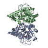 7vglSC 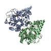 7vgnC 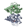 7y5fC 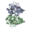 7y5iC 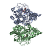 7yheC 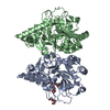 7yw9C S: Starting model for refinement C: citing same article ( |
|---|---|
| Similar structure data |
-Links
 PDBj
PDBj


- Assembly
| Deposited unit | 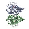
| |||||||||||||||
|---|---|---|---|---|---|---|---|---|---|---|---|---|---|---|---|---|
| 1 |
| |||||||||||||||
| Unit cell |
| |||||||||||||||
| Components on special symmetry positions |
|
-Components
| #1: Protein | Mass: 39087.531 Da / Num. of mol.: 2 Source method: isolated from a genetically manipulated source Source: (gene. exp.) Saccharothrix mutabilis subsp. capreolus (bacteria)Gene: cmnC / Production host: Escherichia coli BL21(DE3) (bacteria) / References: UniProt: A6YEH4#2: Chemical | ChemComp-FE / | Iron  Mass: 55.845 Da / Num. of mol.: 1 / Source method: obtained synthetically / Formula: Fe / Feature type: SUBJECT OF INVESTIGATION Mass: 55.845 Da / Num. of mol.: 1 / Source method: obtained synthetically / Formula: Fe / Feature type: SUBJECT OF INVESTIGATION#3: Chemical | Α-Ketoglutaric acid  Mass: 146.098 Da / Num. of mol.: 2 / Source method: obtained synthetically / Formula: C5H6O5 / Feature type: SUBJECT OF INVESTIGATION Mass: 146.098 Da / Num. of mol.: 2 / Source method: obtained synthetically / Formula: C5H6O5 / Feature type: SUBJECT OF INVESTIGATION#4: Chemical | ChemComp-ARG / | Arginine  Type: L-peptide linking / Mass: 175.209 Da / Num. of mol.: 1 / Source method: obtained synthetically / Formula: C6H15N4O2 / Feature type: SUBJECT OF INVESTIGATION Type: L-peptide linking / Mass: 175.209 Da / Num. of mol.: 1 / Source method: obtained synthetically / Formula: C6H15N4O2 / Feature type: SUBJECT OF INVESTIGATION#5: Water | ChemComp-HOH / | Water Mass: 18.015 Da / Num. of mol.: 503 / Source method: isolated from a natural source / Formula: H2O Mass: 18.015 Da / Num. of mol.: 503 / Source method: isolated from a natural source / Formula: H2OHas ligand of interest | Y | |
|---|
-Experimental details
-Experiment
| Experiment | Method: X-RAY DIFFRACTION / Number of used crystals: 1 |
|---|
- Sample preparation
| Crystal | Density Matthews: 2.78 Å3/Da / Density % sol: 55.72 % |
|---|---|
| Crystal grow | Temperature: 293 K / Method: vapor diffusion, hanging drop / pH: 5.5 Details: 0.2M Ammonium acetate, 0.1M Na citrate tribasic dihydrate pH 5.5, 24% v/v Polyethylene glycol 400 |
-Data collection
| Diffraction | Mean temperature: 100 K / Serial crystal experiment: N |
|---|---|
| Diffraction source | Source: SYNCHROTRON / Site: NSRRC / Beamline: BL15A1 / Wavelength: 0.9732 Å |
| Detector | Type: ADSC QUANTUM 315r / Detector: CCD / Date: Jun 2, 2022 |
| Radiation | Protocol: SINGLE WAVELENGTH / Monochromatic (M) / Laue (L): M / Scattering type: x-ray |
| Radiation wavelength | Wavelength: 0.9732 Å / Relative weight: 1 |
| Reflection | Resolution: 1.7→30 Å / Num. obs: 90120 / % possible obs: 99.7 % / Redundancy: 5.5 % / Rmerge(I) obs: 0.055 / Net I/σ(I): 27.56 |
| Reflection shell | Resolution: 1.7→1.76 Å / Rmerge(I) obs: 0.604 / Mean I/σ(I) obs: 2.42 / Num. unique obs: 8944 / % possible all: 100 |
- Processing
| Software |
| ||||||||||||||||||||||||||||||||||||||||||||||||||||||||||||
|---|---|---|---|---|---|---|---|---|---|---|---|---|---|---|---|---|---|---|---|---|---|---|---|---|---|---|---|---|---|---|---|---|---|---|---|---|---|---|---|---|---|---|---|---|---|---|---|---|---|---|---|---|---|---|---|---|---|---|---|---|---|
| Refinement | Method to determine structure: MOLECULAR REPLACEMENT Starting model: 7VGL Resolution: 1.7→26.28 Å / Cor.coef. Fo:Fc: 0.955 / Cor.coef. Fo:Fc free: 0.939 / SU B: 1.953 / SU ML: 0.063 / Cross valid method: THROUGHOUT / σ(F): 0 / ESU R: 0.097 / ESU R Free: 0.094 / Stereochemistry target values: MAXIMUM LIKELIHOOD Details: HYDROGENS HAVE BEEN ADDED IN THE RIDING POSITIONS U VALUES : REFINED INDIVIDUALLY
| ||||||||||||||||||||||||||||||||||||||||||||||||||||||||||||
| Solvent computation | Ion probe radii: 0.8 Å / Shrinkage radii: 0.8 Å / VDW probe radii: 1.2 Å / Solvent model: MASK | ||||||||||||||||||||||||||||||||||||||||||||||||||||||||||||
| Displacement parameters | Biso max: 76.12 Å2 / Biso mean: 17.297 Å2 / Biso min: 6.52 Å2
| ||||||||||||||||||||||||||||||||||||||||||||||||||||||||||||
| Refinement step | Cycle: final / Resolution: 1.7→26.28 Å
| ||||||||||||||||||||||||||||||||||||||||||||||||||||||||||||
| Refine LS restraints |
| ||||||||||||||||||||||||||||||||||||||||||||||||||||||||||||
| LS refinement shell | Resolution: 1.7→1.744 Å / Rfactor Rfree error: 0 / Total num. of bins used: 20
|
