 Movie
Movie Controller


+ Open data

- Basic information
| Entry | Database: PDB / ID: 7xwg | ||||||||||||
|---|---|---|---|---|---|---|---|---|---|---|---|---|---|
| Title | RSGSGG-AtPRT6 UBR box | ||||||||||||
 Components Components | E3 ubiquitin-protein ligase PRT6 | ||||||||||||
 Keywords Keywords |  LIGASE / PRT6 / UBR box / E3 ubiquitin ligase / Arabidopsis thaliana LIGASE / PRT6 / UBR box / E3 ubiquitin ligase / Arabidopsis thaliana | ||||||||||||
| Function / homology |  Function and homology informationregulation of seed germination / ubiquitin-dependent protein catabolic process via the N-end rule pathway / regulation of lipid catabolic process / response to abscisic acid / defense response to fungus / RING-type E3 ubiquitin transferase / ubiquitin-protein transferase activity / ubiquitin protein ligase activity / ubiquitin-dependent protein catabolic process / protein ubiquitination ...regulation of seed germination / ubiquitin-dependent protein catabolic process via the N-end rule pathway / regulation of lipid catabolic process / response to abscisic acid / defense response to fungus / RING-type E3 ubiquitin transferase / ubiquitin-protein transferase activity / ubiquitin protein ligase activity / ubiquitin-dependent protein catabolic process / protein ubiquitination / defense response to bacterium / zinc ion binding Function and homology informationregulation of seed germination / ubiquitin-dependent protein catabolic process via the N-end rule pathway / regulation of lipid catabolic process / response to abscisic acid / defense response to fungus / RING-type E3 ubiquitin transferase / ubiquitin-protein transferase activity / ubiquitin protein ligase activity / ubiquitin-dependent protein catabolic process / protein ubiquitination ...regulation of seed germination / ubiquitin-dependent protein catabolic process via the N-end rule pathway / regulation of lipid catabolic process / response to abscisic acid / defense response to fungus / RING-type E3 ubiquitin transferase / ubiquitin-protein transferase activity / ubiquitin protein ligase activity / ubiquitin-dependent protein catabolic process / protein ubiquitination / defense response to bacterium / zinc ion bindingSimilarity search - Function | ||||||||||||
| Biological species |  Arabidopsis thaliana (thale cress) Arabidopsis thaliana (thale cress) | ||||||||||||
| Method | X-RAY DIFFRACTION / SYNCHROTRON / MOLECULAR REPLACEMENT / Resolution: 1.832 Å | ||||||||||||
 Authors Authors | Kim, L. / Song, H.K. | ||||||||||||
| Funding support |  Korea, Republic Of, 3items Korea, Republic Of, 3items
| ||||||||||||
 Citation Citation | Journal: To Be Published Title: Structural analyses of plant PRT6-UBR box for Cys-Arg/N-degron pathway and insights into the plant submergence resistance Authors: Kim, L. / Lin, C.C. / Lin, T.J. / Cao, Y.C. / Chen, M.C. / Chou, M.Y. / Lin, W.H. / Kim, M. / Wu, J.L. / Shih, M.C. / Song, H.K. / Ho, M.C. | ||||||||||||
| History |
|
- Structure visualization
| Structure viewer | Molecule: MolmilJmol/JSmol |
|---|
- Downloads & links
-Download
| PDBx/mmCIF format | 7xwg.cif.gz | 60.4 KB | Display | PDBx/mmCIF format |
|---|---|---|---|---|
| PDB format | pdb7xwg.ent.gz | 43.1 KB | Display | PDB format |
| PDBx/mmJSON format | 7xwg.json.gz | Tree view | PDBx/mmJSON format | |
| Others |  Other downloads Other downloads |
-Validation report
| Arichive directory | https://data.pdbj.org/pub/pdb/validation_reports/xw/7xwgftp://data.pdbj.org/pub/pdb/validation_reports/xw/7xwg | HTTPS FTP |
|---|
-Related structure data
| Related structure data |  7xwdC 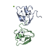 7xweC 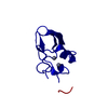 7xwfC  7y6wC 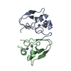 7y6xC 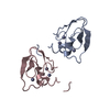 7y6yC  7y6zC  7y70C 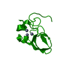 6lhnS S: Starting model for refinement C: citing same article ( |
|---|---|
| Similar structure data |
-Links
 PDBj
PDBj- Assembly
| Deposited unit | 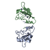
| ||||||||
|---|---|---|---|---|---|---|---|---|---|
| 1 |
| ||||||||
| Unit cell |
|
-Components
| #1: Protein | Mass: 8106.833 Da / Num. of mol.: 2 Source method: isolated from a genetically manipulated source Source: (gene. exp.) Arabidopsis thaliana (thale cress)Gene: PRT6, CER3, GED1, At5g02310/At5g02300, T1E22.70/T1E22.60 Production host:  Escherichia coli (E. coli) Escherichia coli (E. coli)References: UniProt: F4KCC2, RING-type E3 ubiquitin transferase #2: Chemical | ChemComp-ZN /   Mass: 65.409 Da / Num. of mol.: 6 / Source method: obtained synthetically / Formula: Zn / Feature type: SUBJECT OF INVESTIGATION Mass: 65.409 Da / Num. of mol.: 6 / Source method: obtained synthetically / Formula: Zn / Feature type: SUBJECT OF INVESTIGATION#3: Water | ChemComp-HOH / | Water Mass: 18.015 Da / Num. of mol.: 5 / Source method: isolated from a natural source / Formula: H2O Mass: 18.015 Da / Num. of mol.: 5 / Source method: isolated from a natural source / Formula: H2OHas ligand of interest | Y | |
|---|
-Experimental details
-Experiment
| Experiment | Method: X-RAY DIFFRACTION / Number of used crystals: 1 |
|---|
- Sample preparation
| Crystal | Density Matthews: 2.51 Å3/Da / Density % sol: 50.91 % |
|---|---|
| Crystal grow | Temperature: 295 K / Method: vapor diffusion, sitting drop Details: 0.2 M calcium acetate hydrate, 0.1 M sodium cacodylate trihydrate pH 6.5, 18% (w/v) PEG 8000 |
-Data collection
| Diffraction | Mean temperature: 193 K / Serial crystal experiment: N |
|---|---|
| Diffraction source | Source: SYNCHROTRON / Site: PAL/PLS / Beamline: 11C / Wavelength: 1 Å |
| Detector | Type: DECTRIS PILATUS 6M / Detector: PIXEL / Date: Dec 14, 2019 |
| Radiation | Protocol: SINGLE WAVELENGTH / Monochromatic (M) / Laue (L): M / Scattering type: x-ray |
| Radiation wavelength | Wavelength: 1 Å / Relative weight: 1 |
| Reflection | Resolution: 1.832→33.15 Å / Num. obs: 14399 / % possible obs: 97.43 % / Redundancy: 6.6 % / CC1/2: 0.999 / Net I/σ(I): 23 |
| Reflection shell | Resolution: 1.832→1.897 Å / Num. unique obs: 1307 / CC1/2: 0.914 |
- Processing
| Software |
| |||||||||||||||||||||||||||||||||||||||||||||||||||||||||||||||||||||||||||||
|---|---|---|---|---|---|---|---|---|---|---|---|---|---|---|---|---|---|---|---|---|---|---|---|---|---|---|---|---|---|---|---|---|---|---|---|---|---|---|---|---|---|---|---|---|---|---|---|---|---|---|---|---|---|---|---|---|---|---|---|---|---|---|---|---|---|---|---|---|---|---|---|---|---|---|---|---|---|---|
| Refinement | Method to determine structure: MOLECULAR REPLACEMENT Starting model: 6LHN Resolution: 1.832→33.15 Å / SU ML: 0.27 / Cross valid method: FREE R-VALUE / σ(F): 1.34 / Phase error: 34.43 / Stereochemistry target values: ML
| |||||||||||||||||||||||||||||||||||||||||||||||||||||||||||||||||||||||||||||
| Solvent computation | Shrinkage radii: 0.9 Å / VDW probe radii: 1.11 Å / Solvent model: FLAT BULK SOLVENT MODEL | |||||||||||||||||||||||||||||||||||||||||||||||||||||||||||||||||||||||||||||
| Refinement step | Cycle: LAST / Resolution: 1.832→33.15 Å
| |||||||||||||||||||||||||||||||||||||||||||||||||||||||||||||||||||||||||||||
| Refine LS restraints |
| |||||||||||||||||||||||||||||||||||||||||||||||||||||||||||||||||||||||||||||
| LS refinement shell |
|
