 Movie
Movie Controller


+ Open data

- Basic information
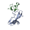
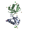
| Entry | Database: PDB / ID: 6lhn | ||||||
|---|---|---|---|---|---|---|---|
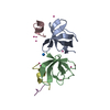
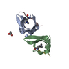
| Title | RLGSGG-AtPRT6 UBR box | ||||||
 Components Components | E3 ubiquitin-protein ligase PRT6 | ||||||
 Keywords Keywords |  LIGASE / PRT6 / Arabidopsis thaliana LIGASE / PRT6 / Arabidopsis thaliana | ||||||
| Function / homology |  Function and homology informationregulation of seed germination / ubiquitin-dependent protein catabolic process via the N-end rule pathway / regulation of lipid catabolic process / response to abscisic acid / defense response to fungus / RING-type E3 ubiquitin transferase / ubiquitin-protein transferase activity / ubiquitin protein ligase activity / ubiquitin-dependent protein catabolic process / protein ubiquitination ...regulation of seed germination / ubiquitin-dependent protein catabolic process via the N-end rule pathway / regulation of lipid catabolic process / response to abscisic acid / defense response to fungus / RING-type E3 ubiquitin transferase / ubiquitin-protein transferase activity / ubiquitin protein ligase activity / ubiquitin-dependent protein catabolic process / protein ubiquitination / defense response to bacterium / zinc ion binding Function and homology informationregulation of seed germination / ubiquitin-dependent protein catabolic process via the N-end rule pathway / regulation of lipid catabolic process / response to abscisic acid / defense response to fungus / RING-type E3 ubiquitin transferase / ubiquitin-protein transferase activity / ubiquitin protein ligase activity / ubiquitin-dependent protein catabolic process / protein ubiquitination ...regulation of seed germination / ubiquitin-dependent protein catabolic process via the N-end rule pathway / regulation of lipid catabolic process / response to abscisic acid / defense response to fungus / RING-type E3 ubiquitin transferase / ubiquitin-protein transferase activity / ubiquitin protein ligase activity / ubiquitin-dependent protein catabolic process / protein ubiquitination / defense response to bacterium / zinc ion bindingSimilarity search - Function | ||||||
| Biological species |  Arabidopsis thaliana (thale cress) Arabidopsis thaliana (thale cress) | ||||||
| Method | X-RAY DIFFRACTION / SYNCHROTRON / MOLECULAR REPLACEMENT / Resolution: 2.5 Å | ||||||
 Authors Authors | Kim, L. / Kwon, D.H. / Song, H.K. | ||||||
 Citation Citation | Journal: J.Biol.Chem. / Year: 2020 Title: Use of the LC3B-fusion technique for biochemical and structural studies of proteins involved in the N-degron pathway. Authors: Kim, L. / Kwon, D.H. / Heo, J. / Park, M.R. / Song, H.K. | ||||||
| History |
|
- Structure visualization
| Structure viewer | Molecule: MolmilJmol/JSmol |
|---|
- Downloads & links
-Download
| PDBx/mmCIF format | 6lhn.cif.gz | 39.6 KB | Display | PDBx/mmCIF format |
|---|---|---|---|---|
| PDB format | pdb6lhn.ent.gz | 26.2 KB | Display | PDB format |
| PDBx/mmJSON format | 6lhn.json.gz | Tree view | PDBx/mmJSON format | |
| Others |  Other downloads Other downloads |
-Validation report
| Arichive directory | https://data.pdbj.org/pub/pdb/validation_reports/lh/6lhnftp://data.pdbj.org/pub/pdb/validation_reports/lh/6lhn | HTTPS FTP |
|---|
-Related structure data
| Related structure data | 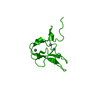 6kgiC 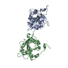 6kgjC  6khzC  6kgg S: Starting model for refinement C: citing same article ( |
|---|---|
| Similar structure data |


-Links
 PDBj
PDBj- Assembly
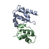
| Deposited unit | 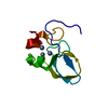
| ||||||||
|---|---|---|---|---|---|---|---|---|---|
| 1 | 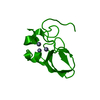
| ||||||||
| Unit cell |
|
-Components
| #1: Protein | Mass: 8189.965 Da / Num. of mol.: 1 / Fragment: UBR box Source method: isolated from a genetically manipulated source Source: (gene. exp.) Arabidopsis thaliana (thale cress)Gene: PRT6, CER3, GED1, At5g02310/At5g02300, T1E22.70/T1E22.60 Production host:  Escherichia coli (E. coli) Escherichia coli (E. coli)References: UniProt: F4KCC2, RING-type E3 ubiquitin transferase | ||
|---|---|---|---|
| #2: Chemical |   Mass: 65.409 Da / Num. of mol.: 3 / Source method: obtained synthetically / Formula: Zn / Feature type: SUBJECT OF INVESTIGATION Mass: 65.409 Da / Num. of mol.: 3 / Source method: obtained synthetically / Formula: Zn / Feature type: SUBJECT OF INVESTIGATIONHas ligand of interest | Y | |
-Experimental details
-Experiment
| Experiment | Method: X-RAY DIFFRACTION / Number of used crystals: 1 |
|---|
- Sample preparation
| Crystal | Density Matthews: 4.45 Å3/Da / Density % sol: 72.36 % |
|---|---|
| Crystal grow | Temperature: 293 K / Method: vapor diffusion, sitting drop / Details: 1.6M ammonium sulfate, 0.1M MES pH6.5 |
-Data collection
| Diffraction | Mean temperature: 193 K / Serial crystal experiment: N |
|---|---|
| Diffraction source | Source: SYNCHROTRON / Site: PAL/PLS  / Beamline: 5C (4A) / Wavelength: 1 Å / Beamline: 5C (4A) / Wavelength: 1 Å |
| Detector | Type: DECTRIS EIGER X 9M / Detector: PIXEL / Date: Nov 24, 2019 |
| Radiation | Protocol: SINGLE WAVELENGTH / Monochromatic (M) / Laue (L): M / Scattering type: x-ray |
| Radiation wavelength | Wavelength: 1 Å / Relative weight: 1 |
| Reflection | Resolution: 2.499→42.67 Å / Num. obs: 5522 / % possible obs: 99.48 % / Redundancy: 9 % / CC1/2: 0.986 / Net I/σ(I): 16.6 |
| Reflection shell | Resolution: 2.5→2.589 Å / Num. unique obs: 5513 / CC1/2: 0.714 |
- Processing
| Software |
| |||||||||||||||||||||||||||||||||||
|---|---|---|---|---|---|---|---|---|---|---|---|---|---|---|---|---|---|---|---|---|---|---|---|---|---|---|---|---|---|---|---|---|---|---|---|---|
| Refinement | Method to determine structure: MOLECULAR REPLACEMENT Starting model: 6KGG 6kgg Resolution: 2.5→38.95 Å / SU ML: 0.27 / Cross valid method: NONE / σ(F): 1.36 / Phase error: 23.4
| |||||||||||||||||||||||||||||||||||
| Solvent computation | Shrinkage radii: 0.9 Å / VDW probe radii: 1.11 Å | |||||||||||||||||||||||||||||||||||
| Refinement step | Cycle: LAST / Resolution: 2.5→38.95 Å
| |||||||||||||||||||||||||||||||||||
| Refine LS restraints |
| |||||||||||||||||||||||||||||||||||
| LS refinement shell |
|
