 Movie
Movie Controller


+ Open data

- Basic information
| Entry | Database: PDB / ID: 7xrh | ||||||
|---|---|---|---|---|---|---|---|
| Title | Feruloyl esterase from Lactobacillus acidophilus | ||||||
 Components Components | Cinnamoyl esterase | ||||||
 Keywords Keywords |  HYDROLASE / Feruloyl esterase HYDROLASE / Feruloyl esterase | ||||||
| Function / homology | Dienelactone hydrolase / Dienelactone hydrolase family / Serine aminopeptidase, S33 / Serine aminopeptidase, S33 / Hydrolases; Acting on ester bonds; Carboxylic-ester hydrolases / aminopeptidase activity / Alpha/Beta hydrolase fold / Cinnamoyl esterase Function and homology information Function and homology information | ||||||
| Biological species |  Lactobacillus acidophilus (bacteria) Lactobacillus acidophilus (bacteria) | ||||||
| Method | X-RAY DIFFRACTION / SYNCHROTRON / MOLECULAR REPLACEMENT / Resolution: 2.3 Å | ||||||
 Authors Authors | Hwang, J. / Lee, C.W. / Lee, J.H. / Do, H. | ||||||
| Funding support |  Korea, Republic Of, 1items Korea, Republic Of, 1items
| ||||||
 Citation Citation | Journal: Int J Mol Sci / Year: 2023 Title: Feruloyl Esterase ( La Fae) from Lactobacillus acidophilus : Structural Insights and Functional Characterization for Application in Ferulic Acid Production. Authors: Jeon, S. / Hwang, J. / Do, H. / Le, L.T.H.L. / Lee, C.W. / Yoo, W. / Lee, M.J. / Shin, S.C. / Kim, K.K. / Kim, H.W. / Lee, J.H. | ||||||
| History |
|
- Structure visualization
| Structure viewer | Molecule: MolmilJmol/JSmol |
|---|
- Downloads & links
-Download
| PDBx/mmCIF format | 7xrh.cif.gz | 104.8 KB | Display | PDBx/mmCIF format |
|---|---|---|---|---|
| PDB format | pdb7xrh.ent.gz | 77.1 KB | Display | PDB format |
| PDBx/mmJSON format | 7xrh.json.gz | Tree view | PDBx/mmJSON format | |
| Others |  Other downloads Other downloads |
-Validation report
| Arichive directory | https://data.pdbj.org/pub/pdb/validation_reports/xr/7xrhftp://data.pdbj.org/pub/pdb/validation_reports/xr/7xrh | HTTPS FTP |
|---|
-Related structure data
| Related structure data | 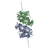 7xriC 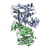 3pf8S S: Starting model for refinement C: citing same article ( |
|---|---|
| Similar structure data |
-Links
 PDBj
PDBj- Assembly
| Deposited unit | 
| ||||||||||||
|---|---|---|---|---|---|---|---|---|---|---|---|---|---|
| 1 |
| ||||||||||||
| Unit cell |
|
-Components
| #1: Protein | Mass: 27487.965 Da / Num. of mol.: 2 Source method: isolated from a genetically manipulated source Details: Several parts of the structure which showed a weak electron density map were deleted. Source: (gene. exp.) Lactobacillus acidophilus (bacteria) / Production host: Escherichia coli (E. coli)References: UniProt: A0A060IN49, Hydrolases; Acting on ester bonds; Carboxylic-ester hydrolases#2: Water | ChemComp-HOH / | Water Mass: 18.015 Da / Num. of mol.: 66 / Source method: isolated from a natural source / Formula: H2O Mass: 18.015 Da / Num. of mol.: 66 / Source method: isolated from a natural source / Formula: H2O |
|---|
-Experimental details
-Experiment
| Experiment | Method: X-RAY DIFFRACTION / Number of used crystals: 1 |
|---|
- Sample preparation
| Crystal | Density Matthews: 2.06 Å3/Da / Density % sol: 40.34 % |
|---|---|
| Crystal grow | Temperature: 293 K / Method: vapor diffusion, sitting drop / pH: 6.5 Details: 20 % (w/v) PEG MME 5000, 0.1 M Bis-Tris-HCl (pH 6.5) |
-Data collection
| Diffraction | Mean temperature: 100 K / Serial crystal experiment: N |
|---|---|
| Diffraction source | Source: SYNCHROTRON / Site: PAL/PLS / Beamline: 5C (4A) / Wavelength: 0.9794 Å |
| Detector | Type: DECTRIS EIGER X 9M / Detector: PIXEL / Date: Dec 9, 2020 |
| Radiation | Protocol: SINGLE WAVELENGTH / Monochromatic (M) / Laue (L): M / Scattering type: x-ray |
| Radiation wavelength | Wavelength: 0.9794 Å / Relative weight: 1 |
| Reflection | Resolution: 2.3→50 Å / Num. obs: 20175 / % possible obs: 97.2 % / Redundancy: 6.8 % / Biso Wilson estimate: 37.4 Å2 / CC1/2: 0.954 / Net I/σ(I): 25.05 |
| Reflection shell | Resolution: 2.3→2.34 Å / Mean I/σ(I) obs: 4.62 / Num. unique obs: 1031 / CC1/2: 0.928 |
- Processing
| Software |
| |||||||||||||||||||||||||||||||||||||||||||||||||||||||||||||||||||||||||||||||||||||||||||||||||||||||||
|---|---|---|---|---|---|---|---|---|---|---|---|---|---|---|---|---|---|---|---|---|---|---|---|---|---|---|---|---|---|---|---|---|---|---|---|---|---|---|---|---|---|---|---|---|---|---|---|---|---|---|---|---|---|---|---|---|---|---|---|---|---|---|---|---|---|---|---|---|---|---|---|---|---|---|---|---|---|---|---|---|---|---|---|---|---|---|---|---|---|---|---|---|---|---|---|---|---|---|---|---|---|---|---|---|---|---|
| Refinement | Method to determine structure: MOLECULAR REPLACEMENT Starting model: 3PF8 Resolution: 2.3→41.06 Å / SU ML: 0.3024 / Cross valid method: FREE R-VALUE / σ(F): 1.34 / Phase error: 30.3203 / Stereochemistry target values: CDL v1.2
| |||||||||||||||||||||||||||||||||||||||||||||||||||||||||||||||||||||||||||||||||||||||||||||||||||||||||
| Solvent computation | Shrinkage radii: 0.9 Å / VDW probe radii: 1.11 Å / Solvent model: FLAT BULK SOLVENT MODEL | |||||||||||||||||||||||||||||||||||||||||||||||||||||||||||||||||||||||||||||||||||||||||||||||||||||||||
| Displacement parameters | Biso mean: 40.02 Å2 | |||||||||||||||||||||||||||||||||||||||||||||||||||||||||||||||||||||||||||||||||||||||||||||||||||||||||
| Refinement step | Cycle: LAST / Resolution: 2.3→41.06 Å
| |||||||||||||||||||||||||||||||||||||||||||||||||||||||||||||||||||||||||||||||||||||||||||||||||||||||||
| Refine LS restraints |
| |||||||||||||||||||||||||||||||||||||||||||||||||||||||||||||||||||||||||||||||||||||||||||||||||||||||||
| LS refinement shell |
|
