 Movie
Movie Controller


+ Open data

- Basic information
| Entry | Database: PDB / ID: 7xds | ||||||
|---|---|---|---|---|---|---|---|
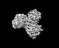
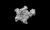
| Title | Crystal structure of wheat stem rust effector AvrSr35 | ||||||
 Components Components | AvrSr35 | ||||||
 Keywords Keywords |  PROTEIN TRANSPORT / Fungus / Effector / Wheat stem rust PROTEIN TRANSPORT / Fungus / Effector / Wheat stem rust | ||||||
| Function / homology | Avirulence factor Function and homology information Function and homology information | ||||||
| Biological species |  Puccinia graminis f. sp. tritici (fungus) Puccinia graminis f. sp. tritici (fungus) | ||||||
| Method | X-RAY DIFFRACTION / SYNCHROTRON / SAD / Resolution: 2.06 Å | ||||||
 Authors Authors | Ouyang, S.Y. / Zhao, Y.B. | ||||||
| Funding support |  China, 1items China, 1items
| ||||||
 Citation Citation | Journal: Sci Adv / Year: 2022 Title: Pathogen effector AvrSr35 triggers Sr35 resistosome assembly via a direct recognition mechanism. Authors: Yan-Bo Zhao / Meng-Xi Liu / Tao-Tao Chen / Xiaomin Ma / Ze-Kai Li / Zichao Zheng / Si-Ru Zheng / Lifei Chen / You-Zhi Li / Li-Rui Tang / Qi Chen / Peiyi Wang / Songying Ouyang / Abstract: Nucleotide-binding, leucine-rich repeat receptors (NLRs) perceive pathogen effectors to trigger plant immunity. The direct recognition mechanism of pathogen effectors by coiled-coil NLRs (CNLs) ...Nucleotide-binding, leucine-rich repeat receptors (NLRs) perceive pathogen effectors to trigger plant immunity. The direct recognition mechanism of pathogen effectors by coiled-coil NLRs (CNLs) remains unclear. We demonstrate that the CNL Sr35 directly recognizes the pathogen effector AvrSr35 from f. sp and report a cryo-electron microscopy structure of Sr35 resistosome and a crystal structure of AvrSr35. We show that AvrSr35 forms homodimers that are disassociated into monomers upon direct recognition by the leucine-rich repeat domain of Sr35, which induces Sr35 resistosome assembly and the subsequent immune response. The first 20 amino-terminal residues of Sr35 are indispensable for immune signaling but not for plasma membrane association. Our findings reveal the direct recognition and activation mechanism of a plant CNL and provide insights into biochemical function of Sr35 resistosome. | ||||||
| History |
|
- Structure visualization
| Structure viewer | Molecule: MolmilJmol/JSmol |
|---|
- Downloads & links
-Download
| PDBx/mmCIF format | 7xds.cif.gz | 177.8 KB | Display | PDBx/mmCIF format |
|---|---|---|---|---|
| PDB format | pdb7xds.ent.gz | 130.4 KB | Display | PDB format |
| PDBx/mmJSON format | 7xds.json.gz | Tree view | PDBx/mmJSON format | |
| Others |  Other downloads Other downloads |
-Validation report
| Arichive directory | https://data.pdbj.org/pub/pdb/validation_reports/xd/7xdsftp://data.pdbj.org/pub/pdb/validation_reports/xd/7xds | HTTPS FTP |
|---|
-Related structure data


-Links
 PDBj
PDBj- Assembly
| Deposited unit | 
| ||||||||||||
|---|---|---|---|---|---|---|---|---|---|---|---|---|---|
| 1 | 
| ||||||||||||
| 2 | 
| ||||||||||||
| Unit cell |
|
-Components
| #1: Protein | Mass: 66621.438 Da / Num. of mol.: 2 Source method: isolated from a genetically manipulated source Source: (gene. exp.) Puccinia graminis f. sp. tritici (fungus)Gene: PGT21_019944, PGTUg99_030428 Production host:  Escherichia coli 'BL21-Gold(DE3)pLysS AG' (bacteria) Escherichia coli 'BL21-Gold(DE3)pLysS AG' (bacteria)References: UniProt: A0A5B0N367 #2: Water | ChemComp-HOH / | Water Mass: 18.015 Da / Num. of mol.: 57 / Source method: isolated from a natural source / Formula: H2O Mass: 18.015 Da / Num. of mol.: 57 / Source method: isolated from a natural source / Formula: H2OHas ligand of interest | Y | |
|---|
-Experimental details
-Experiment
| Experiment | Method: X-RAY DIFFRACTION / Number of used crystals: 1 |
|---|
- Sample preparation
| Crystal grow | Temperature: 289.15 K / Method: vapor diffusion, sitting drop / Details: 0.1 M sodium malonate (pH 6.0), 12% (v/v) PEG 3350 / PH range: 5.6-6.2 |
|---|
-Data collection
| Diffraction | Mean temperature: 100 K / Serial crystal experiment: N |
|---|---|
| Diffraction source | Source: SYNCHROTRON / Site: SSRF / Beamline: BL19U1 / Wavelength: 0.9792 Å |
| Detector | Type: DECTRIS PILATUS3 6M / Detector: PIXEL / Date: Oct 2, 2019 |
| Radiation | Protocol: SINGLE WAVELENGTH / Monochromatic (M) / Laue (L): M / Scattering type: x-ray |
| Radiation wavelength | Wavelength: 0.9792 Å / Relative weight: 1 |
| Reflection | Resolution: 2.06→44.89 Å / Num. obs: 55016 / % possible obs: 97.8 % / Redundancy: 20 % / Biso Wilson estimate: 20.72 Å2 / Rmerge(I) obs: 0.142 / Net I/σ(I): 19 |
| Reflection shell | Resolution: 2.06→2.134 Å / Rmerge(I) obs: 0.142 / Num. unique obs: 55016 |
- Processing
| Software |
| |||||||||||||||||||||||||||||||||||||||||||||||||||||||||||||||||||||||||||||||||||||||||||||||||||||||||||||||||||||||||||||||||||||||||||||||||||
|---|---|---|---|---|---|---|---|---|---|---|---|---|---|---|---|---|---|---|---|---|---|---|---|---|---|---|---|---|---|---|---|---|---|---|---|---|---|---|---|---|---|---|---|---|---|---|---|---|---|---|---|---|---|---|---|---|---|---|---|---|---|---|---|---|---|---|---|---|---|---|---|---|---|---|---|---|---|---|---|---|---|---|---|---|---|---|---|---|---|---|---|---|---|---|---|---|---|---|---|---|---|---|---|---|---|---|---|---|---|---|---|---|---|---|---|---|---|---|---|---|---|---|---|---|---|---|---|---|---|---|---|---|---|---|---|---|---|---|---|---|---|---|---|---|---|---|---|---|
| Refinement | Method to determine structure: SAD / Resolution: 2.06→44.89 Å / SU ML: 0.2834 / Cross valid method: FREE R-VALUE / σ(F): 1.33 / Phase error: 29.374 Stereochemistry target values: GeoStd + Monomer Library + CDL v1.2
| |||||||||||||||||||||||||||||||||||||||||||||||||||||||||||||||||||||||||||||||||||||||||||||||||||||||||||||||||||||||||||||||||||||||||||||||||||
| Solvent computation | Shrinkage radii: 0.9 Å / VDW probe radii: 1.11 Å / Solvent model: FLAT BULK SOLVENT MODEL | |||||||||||||||||||||||||||||||||||||||||||||||||||||||||||||||||||||||||||||||||||||||||||||||||||||||||||||||||||||||||||||||||||||||||||||||||||
| Displacement parameters | Biso mean: 29.13 Å2 | |||||||||||||||||||||||||||||||||||||||||||||||||||||||||||||||||||||||||||||||||||||||||||||||||||||||||||||||||||||||||||||||||||||||||||||||||||
| Refinement step | Cycle: LAST / Resolution: 2.06→44.89 Å
| |||||||||||||||||||||||||||||||||||||||||||||||||||||||||||||||||||||||||||||||||||||||||||||||||||||||||||||||||||||||||||||||||||||||||||||||||||
| Refine LS restraints |
| |||||||||||||||||||||||||||||||||||||||||||||||||||||||||||||||||||||||||||||||||||||||||||||||||||||||||||||||||||||||||||||||||||||||||||||||||||
| LS refinement shell |
|
