 Movie
Movie Controller

[English] 日本語
 Yorodumi
- PDB-7x7n: 3D model of the 3-RBD up single trimeric spike protein of SARS-Co... -
+ Open data

- Basic information
| Entry | Database: PDB / ID: 7x7n | |||||||||
|---|---|---|---|---|---|---|---|---|---|---|
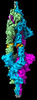
| Title | 3D model of the 3-RBD up single trimeric spike protein of SARS-CoV2 in the presence of synthetic peptide SIH-5. | |||||||||
 Components Components |
| |||||||||
 Keywords Keywords |  VIRAL PROTEIN / spike protein / SARS-CoV2 / complex VIRAL PROTEIN / spike protein / SARS-CoV2 / complex | |||||||||
| Function / homology |  Function and homology information Function and homology informationMaturation of spike protein / viral translation / Translation of Structural Proteins / Virion Assembly and Release / host cell surface / host extracellular space / suppression by virus of host tetherin activity / Induction of Cell-Cell Fusion / structural constituent of virion / host cell endoplasmic reticulum-Golgi intermediate compartment membrane ...Maturation of spike protein / viral translation / Translation of Structural Proteins / Virion Assembly and Release / host cell surface / host extracellular space / suppression by virus of host tetherin activity / Induction of Cell-Cell Fusion / structural constituent of virion / host cell endoplasmic reticulum-Golgi intermediate compartment membrane / entry receptor-mediated virion attachment to host cell / receptor-mediated endocytosis of virus by host cell / Attachment and Entry / membrane fusion / positive regulation of viral entry into host cell / receptor-mediated virion attachment to host cell / receptor ligand activity / host cell surface receptor binding / fusion of virus membrane with host plasma membrane / fusion of virus membrane with host endosome membrane / viral envelope / symbiont-mediated suppression of host type I interferon-mediated signaling pathway / virion attachment to host cell / SARS-CoV-2 activates/modulates innate and adaptive immune responses / host cell plasma membrane / virion membrane / membrane / identical protein binding / plasma membraneSimilarity search - Function | |||||||||
| Biological species |   Severe acute respiratory syndrome coronavirus 2 Severe acute respiratory syndrome coronavirus 2synthetic construct (others) | |||||||||
| Method | ELECTRON MICROSCOPY / single particle reconstruction / cryo EM / Resolution: 4.47 Å | |||||||||
 Authors Authors | Khatri, B. / Pramanick, I. / Malladi, S.K. / Rajmani, R.S. / Kumar, S. / Ghosh, P. / Sengupta, N. / Rahisuddin, R. / Kumaran, S. / Ringe, R.P. ...Khatri, B. / Pramanick, I. / Malladi, S.K. / Rajmani, R.S. / Kumar, S. / Ghosh, P. / Sengupta, N. / Rahisuddin, R. / Kumaran, S. / Ringe, R.P. / Varadarajan, R. / Dutta, S. / Chatterjee, J. | |||||||||
| Funding support |  India, 2items India, 2items
| |||||||||
 Citation Citation | Journal: Nat Chem Biol / Year: 2022 Title: A dimeric proteomimetic prevents SARS-CoV-2 infection by dimerizing the spike protein. Authors: Bhavesh Khatri / Ishika Pramanick / Sameer Kumar Malladi / Raju S Rajmani / Sahil Kumar / Pritha Ghosh / Nayanika Sengupta / R Rahisuddin / Narender Kumar / S Kumaran / Rajesh P Ringe / ...Authors: Bhavesh Khatri / Ishika Pramanick / Sameer Kumar Malladi / Raju S Rajmani / Sahil Kumar / Pritha Ghosh / Nayanika Sengupta / R Rahisuddin / Narender Kumar / S Kumaran / Rajesh P Ringe / Raghavan Varadarajan / Somnath Dutta / Jayanta Chatterjee / Abstract: Protein tertiary structure mimetics are valuable tools to target large protein-protein interaction interfaces. Here, we demonstrate a strategy for designing dimeric helix-hairpin motifs from a ...Protein tertiary structure mimetics are valuable tools to target large protein-protein interaction interfaces. Here, we demonstrate a strategy for designing dimeric helix-hairpin motifs from a previously reported three-helix-bundle miniprotein that targets the receptor-binding domain (RBD) of severe acute respiratory syndrome-coronavirus-2 (SARS-CoV-2). Through truncation of the third helix and optimization of the interhelical loop residues of the miniprotein, we developed a thermostable dimeric helix-hairpin. The dimeric four-helix bundle competes with the human angiotensin-converting enzyme 2 (ACE2) in binding to RBD with 2:2 stoichiometry. Cryogenic-electron microscopy revealed the formation of dimeric spike ectodomain trimer by the four-helix bundle, where all the three RBDs from either spike protein are attached head-to-head in an open conformation, revealing a novel mechanism for virus neutralization. The proteomimetic protects hamsters from high dose viral challenge with replicative SARS-CoV-2 viruses, demonstrating the promise of this class of peptides that inhibit protein-protein interaction through target dimerization. | |||||||||
| History |
|
- Structure visualization
| Structure viewer | Molecule: MolmilJmol/JSmol |
|---|
- Downloads & links
-Download
| PDBx/mmCIF format | 7x7n.cif.gz | 595.3 KB | Display | PDBx/mmCIF format |
|---|---|---|---|---|
| PDB format | pdb7x7n.ent.gz | 488.1 KB | Display | PDB format |
| PDBx/mmJSON format | 7x7n.json.gz | Tree view | PDBx/mmJSON format | |
| Others |  Other downloads Other downloads |
-Validation report
| Arichive directory | https://data.pdbj.org/pub/pdb/validation_reports/x7/7x7nftp://data.pdbj.org/pub/pdb/validation_reports/x7/7x7n | HTTPS FTP |
|---|
-Related structure data
| Related structure data | 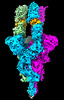 33042MC M: map data used to model this data C: citing same article ( |
|---|---|
| Similar structure data |
-Links
 PDBj
PDBj


- Assembly
| Deposited unit | 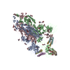
|
|---|---|
| 1 |
|
-Components
| #1: Protein | Spike protein / S glycoprotein / E2 / Peplomer protein Mass: 142399.375 Da / Num. of mol.: 3 / Mutation: R682G, R683S, R685S, K986P, V987P Source method: isolated from a genetically manipulated source Details: NCBI Reference Sequence: YP_009724390.1 In our deposited construct, spike protein is composed of 1-1208 amino acid residues, which have the following mutations R682G, R683S, R685S, K986P, ...Details: NCBI Reference Sequence: YP_009724390.1 In our deposited construct, spike protein is composed of 1-1208 amino acid residues, which have the following mutations R682G, R683S, R685S, K986P, V987P. At the C terminus, there is Foldon oligomerization domain, HRV3C protease site, 6 His, Strep-tag II, linker, Strep-tag II, stop codon. Source: (gene. exp.) Severe acute respiratory syndrome coronavirus 2Gene: S, 2 Cell line (production host): Mammalian expression Expi293F cells Production host:  Homo sapiens (human) / References: UniProt: P0DTC2 Homo sapiens (human) / References: UniProt: P0DTC2#2: Protein/peptide | Mass: 4655.456 Da / Num. of mol.: 6 / Source method: obtained synthetically / Source: (synth.) synthetic construct (others) #3: Sugar | ChemComp-NAG / N-Acetylglucosamine  Type: D-saccharide, beta linking / Mass: 221.208 Da / Num. of mol.: 39 / Source method: obtained synthetically / Formula: C8H15NO6 Type: D-saccharide, beta linking / Mass: 221.208 Da / Num. of mol.: 39 / Source method: obtained synthetically / Formula: C8H15NO6Has ligand of interest | N | |
|---|
-Experimental details
-Experiment
| Experiment | Method: ELECTRON MICROSCOPY |
|---|---|
| EM experiment | Aggregation state: PARTICLE / 3D reconstruction method: single particle reconstruction |
- Sample preparation
| Component |
| ||||||||||||||||||||||||
|---|---|---|---|---|---|---|---|---|---|---|---|---|---|---|---|---|---|---|---|---|---|---|---|---|---|
| Source (natural) | Organism: Severe acute respiratory syndrome coronavirus | ||||||||||||||||||||||||
| Source (recombinant) | Organism: Homo sapiens (human) | ||||||||||||||||||||||||
| Buffer solution | pH: 7.4 | ||||||||||||||||||||||||
| Specimen | Embedding applied: NO / Shadowing applied: NO / Staining applied: NO / Vitrification applied: YES | ||||||||||||||||||||||||
| Vitrification | Cryogen name: ETHANE |
- Electron microscopy imaging
| Experimental equipment |  Model: Talos Arctica / Image courtesy: FEI Company |
|---|---|
| Microscopy | Model: FEI TALOS ARCTICA |
| Electron gun | Electron source: FIELD EMISSION GUN / Accelerating voltage: 200 kV / Illumination mode: OTHER |
| Electron lens | Mode: BRIGHT FIELDBright-field microscopy / Nominal defocus max: 2500 nm / Nominal defocus min: 750 nm |
| Image recording | Electron dose: 80 e/Å2 / Film or detector model: GATAN K2 SUMMIT (4k x 4k) |
- Processing
| Software | Name: PHENIX / Version: 1.17.1_3660: / Classification: refinement | ||||||||||||||||||||||||
|---|---|---|---|---|---|---|---|---|---|---|---|---|---|---|---|---|---|---|---|---|---|---|---|---|---|
| CTF correction | Type: PHASE FLIPPING AND AMPLITUDE CORRECTION | ||||||||||||||||||||||||
| 3D reconstruction | Resolution: 4.47 Å / Resolution method: FSC 0.143 CUT-OFF / Num. of particles: 101296 / Symmetry type: POINT | ||||||||||||||||||||||||
| Refine LS restraints |
|
