 Movie
Movie Controller


+ Open data

- Basic information
| Entry | Database: PDB / ID: 7wvm | |||||||||
|---|---|---|---|---|---|---|---|---|---|---|
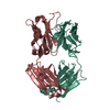
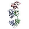
| Title | The complex structure of PD-1 and cemiplimab | |||||||||
 Components Components |
| |||||||||
 Keywords Keywords |  IMMUNE SYSTEM/ANTITUMOR PROTEIN / PD-1 / antibody / N58 glycosylation / cemiplimab / immune checkpoint therapy (ICT) / ANTITUMOR PROTEIN / IMMUNE SYSTEM-ANTITUMOR PROTEIN complex IMMUNE SYSTEM/ANTITUMOR PROTEIN / PD-1 / antibody / N58 glycosylation / cemiplimab / immune checkpoint therapy (ICT) / ANTITUMOR PROTEIN / IMMUNE SYSTEM-ANTITUMOR PROTEIN complex | |||||||||
| Function / homology |  Function and homology information Function and homology informationnegative regulation of tolerance induction / regulatory T cell apoptotic process / negative regulation of immune response / B cell apoptotic process / positive regulation of T cell apoptotic process / negative regulation of B cell apoptotic process / humoral immune response / PD-1 signaling / regulation of immune response / Potential therapeutics for SARS ...negative regulation of tolerance induction / regulatory T cell apoptotic process / negative regulation of immune response / B cell apoptotic process / positive regulation of T cell apoptotic process / negative regulation of B cell apoptotic process / humoral immune response / PD-1 signaling / regulation of immune response / Potential therapeutics for SARS / adaptive immune response / external side of plasma membrane / apoptotic process / plasma membraneSimilarity search - Function | |||||||||
| Biological species |  Homo sapiens (human) Homo sapiens (human) | |||||||||
| Method | X-RAY DIFFRACTION / SYNCHROTRON / MOLECULAR REPLACEMENT / Resolution: 3.4 Å | |||||||||
 Authors Authors | Lu, D. / Xu, Z.P. / Liu, K.F. / Tan, S.G. / Gao, G.F. / Chai, Y. | |||||||||
| Funding support |  China, 2items China, 2items
| |||||||||
 Citation Citation | Journal: Front Immunol / Year: 2022 Title: PD-1 N58-Glycosylation-Dependent Binding of Monoclonal Antibody Cemiplimab for Immune Checkpoint Therapy. Authors: Lu, D. / Xu, Z. / Zhang, D. / Jiang, M. / Liu, K. / He, J. / Ma, D. / Ma, X. / Tan, S. / Gao, G.F. / Chai, Y. | |||||||||
| History |
|
- Structure visualization
| Structure viewer | Molecule: MolmilJmol/JSmol |
|---|
- Downloads & links
-Download
| PDBx/mmCIF format | 7wvm.cif.gz | 151.6 KB | Display | PDBx/mmCIF format |
|---|---|---|---|---|
| PDB format | pdb7wvm.ent.gz | 108.8 KB | Display | PDB format |
| PDBx/mmJSON format | 7wvm.json.gz | Tree view | PDBx/mmJSON format | |
| Others |  Other downloads Other downloads |
-Validation report
| Arichive directory | https://data.pdbj.org/pub/pdb/validation_reports/wv/7wvmftp://data.pdbj.org/pub/pdb/validation_reports/wv/7wvm | HTTPS FTP |
|---|
-Related structure data
-Links
 PDBj
PDBj


- Assembly
| Deposited unit | 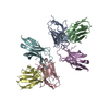
| ||||||||||||
|---|---|---|---|---|---|---|---|---|---|---|---|---|---|
| 1 | 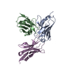
| ||||||||||||
| 2 | 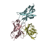
| ||||||||||||
| Unit cell |
|
-Components
| #1: Antibody | Mass: 12751.239 Da / Num. of mol.: 2 Source method: isolated from a genetically manipulated source Source: (gene. exp.) Homo sapiens (human) / Production host:  Escherichia coli (E. coli) Escherichia coli (E. coli)#2: Antibody | Mass: 11514.724 Da / Num. of mol.: 2 Source method: isolated from a genetically manipulated source Source: (gene. exp.) Homo sapiens (human) / Production host: Escherichia coli (E. coli)#3: Protein | / Protein PD-1 / hPD-1Mass: 13170.722 Da / Num. of mol.: 2 Source method: isolated from a genetically manipulated source Source: (gene. exp.) Homo sapiens (human) / Gene: PDCD1, PD1 / Production host: Escherichia coli (E. coli) / References: UniProt: Q15116 |
|---|
-Experimental details
-Experiment
| Experiment | Method: X-RAY DIFFRACTION / Number of used crystals: 1 |
|---|
- Sample preparation
| Crystal | Density Matthews: 4.22 Å3/Da / Density % sol: 70.84 % |
|---|---|
| Crystal grow | Temperature: 291 K / Method: vapor diffusion, sitting drop Details: 0.1 M Sodium acetate, pH 5.0, 5% w/v PGA (Na+ form, LM), 20% w/v PEG 2000 MME |
-Data collection
| Diffraction | Mean temperature: 100 K / Serial crystal experiment: N |
|---|---|
| Diffraction source | Source: SYNCHROTRON / Site: SSRF / Beamline: BL19U1 / Wavelength: 0.97853 Å |
| Detector | Type: DECTRIS PILATUS 6M / Detector: PIXEL / Date: Jan 15, 2021 |
| Radiation | Protocol: SINGLE WAVELENGTH / Monochromatic (M) / Laue (L): M / Scattering type: x-ray |
| Radiation wavelength | Wavelength: 0.97853 Å / Relative weight: 1 |
| Reflection | Resolution: 3.4→50 Å / Num. obs: 17566 / % possible obs: 100 % / Redundancy: 8.9 % / Biso Wilson estimate: 68.82 Å2 / Rmerge(I) obs: 0.2 / Net I/σ(I): 9.5 |
| Reflection shell | Resolution: 3.4→3.52 Å / Rmerge(I) obs: 0.658 / Num. unique obs: 1721 |
- Processing
| Software |
| |||||||||||||||||||||||||||||||||||||||||||||||||
|---|---|---|---|---|---|---|---|---|---|---|---|---|---|---|---|---|---|---|---|---|---|---|---|---|---|---|---|---|---|---|---|---|---|---|---|---|---|---|---|---|---|---|---|---|---|---|---|---|---|---|
| Refinement | Method to determine structure: MOLECULAR REPLACEMENT Starting model: 5GGU, 6KTR Resolution: 3.4→43.06 Å / SU ML: 0.4753 / Cross valid method: FREE R-VALUE / σ(F): 1.34 / Phase error: 33.151 Stereochemistry target values: GeoStd + Monomer Library + CDL v1.2
| |||||||||||||||||||||||||||||||||||||||||||||||||
| Solvent computation | Shrinkage radii: 0.9 Å / VDW probe radii: 1.11 Å / Solvent model: FLAT BULK SOLVENT MODEL | |||||||||||||||||||||||||||||||||||||||||||||||||
| Displacement parameters | Biso mean: 63.12 Å2 | |||||||||||||||||||||||||||||||||||||||||||||||||
| Refinement step | Cycle: LAST / Resolution: 3.4→43.06 Å
| |||||||||||||||||||||||||||||||||||||||||||||||||
| Refine LS restraints |
| |||||||||||||||||||||||||||||||||||||||||||||||||
| LS refinement shell |
|
