 Movie
Movie Controller

[English] 日本語
 Yorodumi
- PDB-7wbr: Citrate synthase/lyase from Desulfurella acetivorans Desace_08345 -
+ Open data

- Basic information
| Entry | Database: PDB / ID: 7wbr | ||||||
|---|---|---|---|---|---|---|---|
| Title | Citrate synthase/lyase from Desulfurella acetivorans Desace_08345 | ||||||
 Components Components | Citrate synthase | ||||||
 Keywords Keywords | TRANSFERASE / Citrate synthase / probable citrate lyase / TCA / roTCA | ||||||
| Function / homology |  Function and homology information Function and homology informationacyltransferase activity, acyl groups converted into alkyl on transfer / citrate synthase (unknown stereospecificity) / tricarboxylic acid cycleSimilarity search - Function | ||||||
| Biological species |  Desulfurella acetivorans (bacteria) Desulfurella acetivorans (bacteria) | ||||||
| Method | X-RAY DIFFRACTION / SYNCHROTRON / MOLECULAR REPLACEMENT / Resolution: 2.19 Å | ||||||
 Authors Authors | Yang, L. | ||||||
| Funding support |  China, 1items China, 1items
| ||||||
 Citation Citation | Journal: To Be Published Title: Structure of Citrate synthase/lyase from Desulfurella acetivorans Desace_08345 complex with citrate Authors: Yang, L. | ||||||
| History |
|
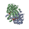
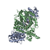
- Structure visualization
| Structure viewer | Molecule: MolmilJmol/JSmol |
|---|
- Downloads & links
-Download
| PDBx/mmCIF format | 7wbr.cif.gz | 663.3 KB | Display | PDBx/mmCIF format |
|---|---|---|---|---|
| PDB format | pdb7wbr.ent.gz | 551 KB | Display | PDB format |
| PDBx/mmJSON format | 7wbr.json.gz | Tree view | PDBx/mmJSON format | |
| Others |  Other downloads Other downloads |
-Validation report
| Arichive directory | https://data.pdbj.org/pub/pdb/validation_reports/wb/7wbrftp://data.pdbj.org/pub/pdb/validation_reports/wb/7wbr | HTTPS FTP |
|---|
-Related structure data
| Related structure data |  1ixeS S: Starting model for refinement |
|---|---|
| Similar structure data |


-Links
 PDBj
PDBj

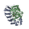
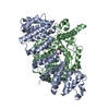
- Assembly
| Deposited unit | 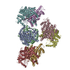
| ||||||||
|---|---|---|---|---|---|---|---|---|---|
| 1 | 
| ||||||||
| 2 | 
| ||||||||
| 3 | 
| ||||||||
| 4 | 
| ||||||||
| Unit cell |
|
-Components
| #1: Protein | / lyase Desace_08345 Mass: 49056.430 Da / Num. of mol.: 8 / Mutation: L3F/C48S/I99V/V207L/T278V/A309S/M330V Source method: isolated from a genetically manipulated source Source: (gene. exp.) Desulfurella acetivorans (bacteria) / Gene: ENO40_01140 / Production host:  Escherichia phage EcSzw-2 (virus) Escherichia phage EcSzw-2 (virus)References: UniProt: A0A7C2VVZ8, citrate synthase (unknown stereospecificity) #2: Chemical | ChemComp-CIT / Citric acid  Mass: 192.124 Da / Num. of mol.: 8 / Source method: obtained synthetically / Formula: C6H8O7 / Feature type: SUBJECT OF INVESTIGATION Mass: 192.124 Da / Num. of mol.: 8 / Source method: obtained synthetically / Formula: C6H8O7 / Feature type: SUBJECT OF INVESTIGATION#3: Water | ChemComp-HOH / | Water Mass: 18.015 Da / Num. of mol.: 973 / Source method: isolated from a natural source / Formula: H2O Mass: 18.015 Da / Num. of mol.: 973 / Source method: isolated from a natural source / Formula: H2OHas ligand of interest | Y | |
|---|
-Experimental details
-Experiment
| Experiment | Method: X-RAY DIFFRACTION / Number of used crystals: 1 |
|---|
- Sample preparation
| Crystal | Density Matthews: 2.57 Å3/Da / Density % sol: 52.11 % |
|---|---|
| Crystal grow | Temperature: 289.15 K / Method: vapor diffusion, sitting drop / Details: PEG 3350, Ammonium citrate tribasic pH 7.0 |
-Data collection
| Diffraction | Mean temperature: 100 K / Serial crystal experiment: N |
|---|---|
| Diffraction source | Source: SYNCHROTRON / Site: SSRF / Beamline: BL18U1 / Wavelength: 0.97915 Å |
| Detector | Type: DECTRIS EIGER X 4M / Detector: PIXEL / Date: Dec 8, 2021 |
| Radiation | Protocol: SINGLE WAVELENGTH / Monochromatic (M) / Laue (L): M / Scattering type: x-ray |
| Radiation wavelength | Wavelength: 0.97915 Å / Relative weight: 1 |
| Reflection | Resolution: 2.18→181.17 Å / Num. obs: 191541 / % possible obs: 97.5 % / Redundancy: 8.2 % / CC1/2: 0.991 / Rmerge(I) obs: 0.129 / Net I/σ(I): 32 |
| Reflection shell | Resolution: 2.18→2.22 Å / Redundancy: 8 % / Rmerge(I) obs: 0.567 / Num. unique obs: 9494 / CC star: 0.964 / Rpim(I) all: 0.21 / Rrim(I) all: 0.606 |
- Processing
| Software |
| ||||||||||||||||||||||||||||||||||||||||||||||||||||||||||||
|---|---|---|---|---|---|---|---|---|---|---|---|---|---|---|---|---|---|---|---|---|---|---|---|---|---|---|---|---|---|---|---|---|---|---|---|---|---|---|---|---|---|---|---|---|---|---|---|---|---|---|---|---|---|---|---|---|---|---|---|---|---|
| Refinement | Method to determine structure: MOLECULAR REPLACEMENT Starting model: 1ixe Resolution: 2.19→181.17 Å / Cor.coef. Fo:Fc: 0.946 / Cor.coef. Fo:Fc free: 0.912 / SU B: 6.415 / SU ML: 0.163 / Cross valid method: THROUGHOUT / σ(F): 0 / ESU R: 0.273 / ESU R Free: 0.218 / Stereochemistry target values: MAXIMUM LIKELIHOOD Details: HYDROGENS HAVE BEEN ADDED IN THE RIDING POSITIONS U VALUES : REFINED INDIVIDUALLY
| ||||||||||||||||||||||||||||||||||||||||||||||||||||||||||||
| Solvent computation | Ion probe radii: 0.8 Å / Shrinkage radii: 0.8 Å / VDW probe radii: 1.2 Å / Solvent model: MASK | ||||||||||||||||||||||||||||||||||||||||||||||||||||||||||||
| Displacement parameters | Biso max: 140.01 Å2 / Biso mean: 36.133 Å2 / Biso min: 13.36 Å2
| ||||||||||||||||||||||||||||||||||||||||||||||||||||||||||||
| Refinement step | Cycle: final / Resolution: 2.19→181.17 Å
| ||||||||||||||||||||||||||||||||||||||||||||||||||||||||||||
| Refine LS restraints |
| ||||||||||||||||||||||||||||||||||||||||||||||||||||||||||||
| LS refinement shell | Resolution: 2.19→2.245 Å / Rfactor Rfree error: 0
|
