 Movie
Movie Controller

[English] 日本語
 Yorodumi
- PDB-7viy: class II photolyase MmCPDII oxidized to semiquinone TR-SFX studie... -
+ Open data

- Basic information
| Entry | Database: PDB / ID: 7viy | |||||||||
|---|---|---|---|---|---|---|---|---|---|---|
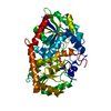
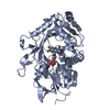
| Title | class II photolyase MmCPDII oxidized to semiquinone TR-SFX studies (50 ns time-point) | |||||||||
 Components Components | DNA photolyase | |||||||||
 Keywords Keywords |  FLAVOPROTEIN / photolyase / electron transport / photoreduction / time-resolved serial crystallography FLAVOPROTEIN / photolyase / electron transport / photoreduction / time-resolved serial crystallography | |||||||||
| Function / homology |  Function and homology information Function and homology informationphotoreactive repair / deoxyribodipyrimidine photo-lyase / deoxyribodipyrimidine photo-lyase activity / nucleotide binding / DNA bindingSimilarity search - Function | |||||||||
| Biological species |  Methanosarcina mazei Go1 (archaea) Methanosarcina mazei Go1 (archaea) | |||||||||
| Method | X-RAY DIFFRACTION / FREE ELECTRON LASER / MOLECULAR REPLACEMENT / Resolution: 2.25 Å | |||||||||
 Authors Authors | Maestre-Reyna, M. / Yang, C.-H. / Huang, W.-C. / Nango, E. / Ngura Putu, E.P.G. / Franz-Badur, S. / Wu, W.-J. / Wu, H.-Y. / Wang, P.-H. / Hosokawa, Y. ...Maestre-Reyna, M. / Yang, C.-H. / Huang, W.-C. / Nango, E. / Ngura Putu, E.P.G. / Franz-Badur, S. / Wu, W.-J. / Wu, H.-Y. / Wang, P.-H. / Hosokawa, Y. / Saft, M. / Emmerich, H.-J. / Liao, J.-H. / Lee, C.-C. / Huang, K.-F. / Chang, Y.-K. / Weng, J.-H. / Royant, A. / Gad, W. / Pang, A.H. / Chang, C.-W. / Sugahara, M. / Owada, S. / Joti, Y. / Yamashita, A. / Tanaka, R. / Tanaka, T. / Luo, F.J. / Tono, K. / Kiontke, S. / Yamamoto, J. / Iwata, S. / Essen, L.-O. / Bessho, Y. / Tsai, M.-D. | |||||||||
| Funding support |  Japan, 2items Japan, 2items
| |||||||||
 Citation Citation | Journal: Nat.Chem. / Year: 2022 Title: Serial crystallography captures dynamic control of sequential electron and proton transfer events in a flavoenzyme. Authors: Maestre-Reyna, M. / Yang, C.H. / Nango, E. / Huang, W.C. / Ngurah Putu, E.P.G. / Wu, W.J. / Wang, P.H. / Franz-Badur, S. / Saft, M. / Emmerich, H.J. / Wu, H.Y. / Lee, C.C. / Huang, K.F. / ...Authors: Maestre-Reyna, M. / Yang, C.H. / Nango, E. / Huang, W.C. / Ngurah Putu, E.P.G. / Wu, W.J. / Wang, P.H. / Franz-Badur, S. / Saft, M. / Emmerich, H.J. / Wu, H.Y. / Lee, C.C. / Huang, K.F. / Chang, Y.K. / Liao, J.H. / Weng, J.H. / Gad, W. / Chang, C.W. / Pang, A.H. / Sugahara, M. / Owada, S. / Hosokawa, Y. / Joti, Y. / Yamashita, A. / Tanaka, R. / Tanaka, T. / Luo, F. / Tono, K. / Hsu, K.C. / Kiontke, S. / Schapiro, I. / Spadaccini, R. / Royant, A. / Yamamoto, J. / Iwata, S. / Essen, L.O. / Bessho, Y. / Tsai, M.D. | |||||||||
| History |
|
- Structure visualization
| Structure viewer | Molecule: MolmilJmol/JSmol |
|---|
- Downloads & links
-Download
| PDBx/mmCIF format | 7viy.cif.gz | 203 KB | Display | PDBx/mmCIF format |
|---|---|---|---|---|
| PDB format | pdb7viy.ent.gz | 157.5 KB | Display | PDB format |
| PDBx/mmJSON format | 7viy.json.gz | Tree view | PDBx/mmJSON format | |
| Others |  Other downloads Other downloads |
-Validation report
| Arichive directory | https://data.pdbj.org/pub/pdb/validation_reports/vi/7viyftp://data.pdbj.org/pub/pdb/validation_reports/vi/7viy | HTTPS FTP |
|---|
-Related structure data
| Related structure data | 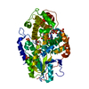 7f8tC 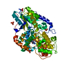 7viwC 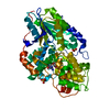 7vixC  7vizC 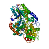 7vj0C 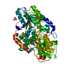 7vj1C 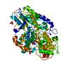 7vj2C 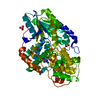 7vj3C 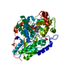 7vj4C  7vj5C  7vj6C  7vj7C  7vj8C  7vj9C  7vjaC 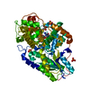 7vjbC 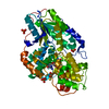 7vjcC  7vjeC 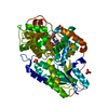 7vjgC 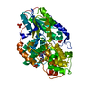 7vjhC  7vjiC 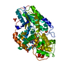 7vjjC 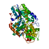 7vjkC  6lm4 C: citing same article ( S: Starting model for refinement |
|---|---|
| Similar structure data |


-Links
 PDBj
PDBj
- Assembly
| Deposited unit | 
| ||||||||
|---|---|---|---|---|---|---|---|---|---|
| 1 |
| ||||||||
| Unit cell |
|
-Components
| #1: Protein | Mass: 55123.480 Da / Num. of mol.: 1 / Mutation: M377T Source method: isolated from a genetically manipulated source Source: (gene. exp.) Methanosarcina mazei Go1 (archaea) / Strain: Go1 / Gene: MM_0852 / Production host:  Escherichia coli BL21(DE3) (bacteria) / Strain (production host): BL21(DE3) Escherichia coli BL21(DE3) (bacteria) / Strain (production host): BL21(DE3)References: UniProt: Q8PYK9, deoxyribodipyrimidine photo-lyase |
|---|---|
| #2: Chemical | ChemComp-SO4 / Sulfate  Mass: 96.063 Da / Num. of mol.: 1 / Source method: obtained synthetically / Formula: SO4 Mass: 96.063 Da / Num. of mol.: 1 / Source method: obtained synthetically / Formula: SO4 |
| #3: Chemical | ChemComp-FAD / Flavin adenine dinucleotide  Mass: 785.550 Da / Num. of mol.: 1 / Source method: obtained synthetically / Formula: C27H33N9O15P2 / Feature type: SUBJECT OF INVESTIGATION / Comment: FAD*YM Mass: 785.550 Da / Num. of mol.: 1 / Source method: obtained synthetically / Formula: C27H33N9O15P2 / Feature type: SUBJECT OF INVESTIGATION / Comment: FAD*YM |
| #4: Water | ChemComp-HOH / Water Mass: 18.015 Da / Num. of mol.: 132 / Source method: isolated from a natural source / Formula: H2O Mass: 18.015 Da / Num. of mol.: 132 / Source method: isolated from a natural source / Formula: H2O |
| Has ligand of interest | Y |
-Experimental details
-Experiment
| Experiment | Method: X-RAY DIFFRACTION / Number of used crystals: 1 |
|---|
- Sample preparation
| Crystal | Density Matthews: 2.8 Å3/Da / Density % sol: 56.12 % |
|---|---|
| Crystal grow | Temperature: 278 K / Method: batch mode / Details: 0.5-0.65 M Li2SO4 10-15% (W/V) PEG8000 |
-Data collection
| Diffraction | Mean temperature: 293 K / Serial crystal experiment: Y |
|---|---|
| Diffraction source | Source: FREE ELECTRON LASER / Site: SACLA / Beamline: BL2 / Wavelength: 1.2398 Å |
| Detector | Type: MPCCD / Detector: CCD / Date: May 28, 2019 |
| Radiation | Protocol: SINGLE WAVELENGTH / Monochromatic (M) / Laue (L): M / Scattering type: x-ray |
| Radiation wavelength | Wavelength: 1.2398 Å / Relative weight: 1 |
| Reflection | Resolution: 1.95→35.57 Å / Num. obs: 47026 / % possible obs: 100 % / Redundancy: 805.8 % / CC1/2: 0.997 / Net I/σ(I): 8 |
| Reflection shell | Resolution: 1.95→1.97 Å / Mean I/σ(I) obs: 0.67 / Num. unique obs: 2278 / CC1/2: 0.39 / % possible all: 100 |
| Serial crystallography measurement | Focal spot size: 1.77 µm2 / Pulse duration: 10 fsec. / Pulse photon energy: 10 keV / XFEL pulse repetition rate: 30 Hz |
| Serial crystallography sample delivery | Description: high viscosity injector / Method: injection |
| Serial crystallography sample delivery injection | Carrier solvent: grease / Filter size: 70 µm / Flow rate: 4.5 µL/min / Injector diameter: 100 µm / Injector temperature: 293 K / Jet diameter: 100 µm / Power by: gas |
| Serial crystallography data reduction | Crystal hits: 39644 |
- Processing
| Software |
| ||||||||||||||||||||||||||||||||||||||||||||||||||||||||||||||||||||||||||||||||||||
|---|---|---|---|---|---|---|---|---|---|---|---|---|---|---|---|---|---|---|---|---|---|---|---|---|---|---|---|---|---|---|---|---|---|---|---|---|---|---|---|---|---|---|---|---|---|---|---|---|---|---|---|---|---|---|---|---|---|---|---|---|---|---|---|---|---|---|---|---|---|---|---|---|---|---|---|---|---|---|---|---|---|---|---|---|---|
| Refinement | Method to determine structure: MOLECULAR REPLACEMENT Starting model: 6LM4 6lm4 Resolution: 2.25→35.57 Å / SU ML: 0.38 / Cross valid method: THROUGHOUT / σ(F): 0 / Phase error: 33.43 / Stereochemistry target values: ML
| ||||||||||||||||||||||||||||||||||||||||||||||||||||||||||||||||||||||||||||||||||||
| Solvent computation | Shrinkage radii: 0.9 Å / VDW probe radii: 1.11 Å / Solvent model: FLAT BULK SOLVENT MODEL | ||||||||||||||||||||||||||||||||||||||||||||||||||||||||||||||||||||||||||||||||||||
| Refinement step | Cycle: LAST / Resolution: 2.25→35.57 Å
| ||||||||||||||||||||||||||||||||||||||||||||||||||||||||||||||||||||||||||||||||||||
| Refine LS restraints |
| ||||||||||||||||||||||||||||||||||||||||||||||||||||||||||||||||||||||||||||||||||||
| LS refinement shell |
| ||||||||||||||||||||||||||||||||||||||||||||||||||||||||||||||||||||||||||||||||||||
| Refinement TLS params. | Method: refined / Origin x: 19.6021 Å / Origin y: 24.9629 Å / Origin z: 106.4069 Å
| ||||||||||||||||||||||||||||||||||||||||||||||||||||||||||||||||||||||||||||||||||||
| Refinement TLS group | Selection details: all |
