 Movie
Movie Controller

[English] 日本語
 Yorodumi
- PDB-5oc2: Crystal structure of Asp295Cys/Lys303Cys Amadoriase I mutant from... -
+ Open data

- Basic information
| Entry | Database: PDB / ID: 5oc2 | ||||||
|---|---|---|---|---|---|---|---|
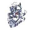
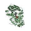
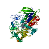
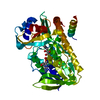
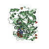
| Title | Crystal structure of Asp295Cys/Lys303Cys Amadoriase I mutant from Aspergillus Fumigatus | ||||||
 Components Components | Fructosyl amine:oxygen oxidoreductase | ||||||
 Keywords Keywords |  OXIDOREDUCTASE / thermoresistance / flavin dependant enzyme / glycated aminoacid OXIDOREDUCTASE / thermoresistance / flavin dependant enzyme / glycated aminoacid | ||||||
| Function / homology |  Function and homology information Function and homology informationfructosyl-amino acid oxidase activity / saccharopine oxidase activity / sarcosine oxidase activity / flavin adenine dinucleotide binding / oxidoreductase activitySimilarity search - Function | ||||||
| Biological species |  Neosartorya fumigata (mold) Neosartorya fumigata (mold) | ||||||
| Method | X-RAY DIFFRACTION / SYNCHROTRON / MOLECULAR REPLACEMENT / Resolution: 2.85 Å | ||||||
 Authors Authors | Rigoldi, F. / Donini, S. / Gautieri, A. / Parisini, E. | ||||||
| Funding support |  Italy, 1items Italy, 1items
| ||||||
 Citation Citation | Journal: Sci Rep / Year: 2018 Title: Thermal stabilization of the deglycating enzyme Amadoriase I by rational design. Authors: Rigoldi, F. / Donini, S. / Giacomina, F. / Sorana, F. / Redaelli, A. / Bandiera, T. / Parisini, E. / Gautieri, A. | ||||||
| History |
|
- Structure visualization
| Structure viewer | Molecule: MolmilJmol/JSmol |
|---|
- Downloads & links
-Download
| PDBx/mmCIF format | 5oc2.cif.gz | 193 KB | Display | PDBx/mmCIF format |
|---|---|---|---|---|
| PDB format | pdb5oc2.ent.gz | 150.7 KB | Display | PDB format |
| PDBx/mmJSON format | 5oc2.json.gz | Tree view | PDBx/mmJSON format | |
| Others |  Other downloads Other downloads |
-Validation report
| Arichive directory | https://data.pdbj.org/pub/pdb/validation_reports/oc/5oc2ftp://data.pdbj.org/pub/pdb/validation_reports/oc/5oc2 | HTTPS FTP |
|---|
-Related structure data
| Related structure data |  5oc3C  4wctS S: Starting model for refinement C: citing same article ( |
|---|---|
| Similar structure data |

-Links
 PDBj
PDBj- Assembly
| Deposited unit | 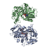
| ||||||||
|---|---|---|---|---|---|---|---|---|---|
| 1 | 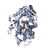
| ||||||||
| 2 | 
| ||||||||
| Unit cell |
|
-Components
| #1: Protein | Mass: 51298.766 Da / Num. of mol.: 2 Source method: isolated from a genetically manipulated source Source: (gene. exp.) Neosartorya fumigata (mold)Production host:  Escherichia coli 'BL21-Gold(DE3)pLysS AG' (bacteria) Escherichia coli 'BL21-Gold(DE3)pLysS AG' (bacteria)References: UniProt: O42629, UniProt: Q4WIF5*PLUS #2: Chemical | Flavin adenine dinucleotide  Mass: 785.550 Da / Num. of mol.: 2 / Source method: obtained synthetically / Formula: C27H33N9O15P2 / Comment: FAD*YM Mass: 785.550 Da / Num. of mol.: 2 / Source method: obtained synthetically / Formula: C27H33N9O15P2 / Comment: FAD*YM#3: Water | ChemComp-HOH / | Water Mass: 18.015 Da / Num. of mol.: 282 / Source method: isolated from a natural source / Formula: H2O Mass: 18.015 Da / Num. of mol.: 282 / Source method: isolated from a natural source / Formula: H2O |
|---|
-Experimental details
-Experiment
| Experiment | Method: X-RAY DIFFRACTION / Number of used crystals: 1 |
|---|
- Sample preparation
| Crystal | Density Matthews: 2.41 Å3/Da / Density % sol: 48.87 % |
|---|---|
| Crystal grow | Temperature: 298 K / Method: vapor diffusion, hanging drop Details: 0.1 M sodium citrate pH 5.6 14% Peg4K 5 % dimethyl sulfoxide |
-Data collection
| Diffraction | Mean temperature: 100 K |
|---|---|
| Diffraction source | Source: SYNCHROTRON / Site: SLS  / Beamline: X06DA / Wavelength: 1 Å / Beamline: X06DA / Wavelength: 1 Å |
| Detector | Type: PSI PILATUS 6M / Detector: PIXEL / Date: May 29, 2017 |
| Radiation | Protocol: SINGLE WAVELENGTH / Monochromatic (M) / Laue (L): M / Scattering type: x-ray |
| Radiation wavelength | Wavelength: 1 Å / Relative weight: 1 |
| Reflection | Resolution: 2.85→48.82 Å / Num. obs: 22860 / % possible obs: 100 % / Redundancy: 6.9 % / Rmerge(I) obs: 0.165 / Net I/av σ(I): 10.6 / Net I/σ(I): 10.59 |
| Reflection shell | Resolution: 2.85→3 Å / Redundancy: 6.6 % / Rmerge(I) obs: 0.495 / Mean I/σ(I) obs: 4.1 / Num. unique obs: 3316 / % possible all: 100 |
- Processing
| Software |
| |||||||||||||||||||||||||||||||||||||||||||||||||||||||||||||||
|---|---|---|---|---|---|---|---|---|---|---|---|---|---|---|---|---|---|---|---|---|---|---|---|---|---|---|---|---|---|---|---|---|---|---|---|---|---|---|---|---|---|---|---|---|---|---|---|---|---|---|---|---|---|---|---|---|---|---|---|---|---|---|---|---|
| Refinement | Method to determine structure: MOLECULAR REPLACEMENT Starting model: 4wct Resolution: 2.85→46.371 Å / SU ML: 0.34 / Cross valid method: FREE R-VALUE / σ(F): 1.34 / Phase error: 25.17 / Stereochemistry target values: ML
| |||||||||||||||||||||||||||||||||||||||||||||||||||||||||||||||
| Solvent computation | Shrinkage radii: 0.9 Å / VDW probe radii: 1.11 Å / Solvent model: FLAT BULK SOLVENT MODEL | |||||||||||||||||||||||||||||||||||||||||||||||||||||||||||||||
| Refinement step | Cycle: LAST / Resolution: 2.85→46.371 Å
| |||||||||||||||||||||||||||||||||||||||||||||||||||||||||||||||
| Refine LS restraints |
| |||||||||||||||||||||||||||||||||||||||||||||||||||||||||||||||
| LS refinement shell |
|
