 Movie
Movie Controller

[English] 日本語
 Yorodumi
- PDB-7ve2: Crystal Structure of Lopinavir bound Plasmepsin II (PMII) from Pl... -
+ Open data

- Basic information
| Entry | Database: PDB / ID: 7ve2 | ||||||
|---|---|---|---|---|---|---|---|
| Title | Crystal Structure of Lopinavir bound Plasmepsin II (PMII) from Plasmodium falciparum | ||||||
 Components Components | Plasmepsin II | ||||||
 Keywords Keywords | HYDROLASE / Inhibitor / Protease / Peptidomimetic / HIV-1 protease inhibitor / peptidase / hemoglobin degrader | ||||||
| Function / homology |  Function and homology information Function and homology informationMHC class II antigen presentation / hemoglobin catabolic process / cytostome / plasmepsin II / Neutrophil degranulation / vacuolar lumen / food vacuole / vacuolar membrane / aspartic-type endopeptidase activity / proteolysisSimilarity search - Function | ||||||
| Biological species |  Plasmodium falciparum (malaria parasite P. falciparum) Plasmodium falciparum (malaria parasite P. falciparum) | ||||||
| Method | X-RAY DIFFRACTION / MOLECULAR REPLACEMENT / Resolution: 3.2 Å | ||||||
 Authors Authors | Mishra, V. / Rathore, I. / Bhaumik, P. | ||||||
| Funding support |  India, 1items India, 1items
| ||||||
 Citation Citation | Journal: Curr Res Struct Biol / Year: 2024 Title: Inhibition of Plasmodium falciparum plasmepsins by drugs targeting HIV-1 protease: A way forward for antimalarial drug discovery. Authors: Mishra, V. / Deshmukh, A. / Rathore, I. / Chakraborty, S. / Patankar, S. / Gustchina, A. / Wlodawer, A. / Yada, R.Y. / Bhaumik, P. #1: Journal: Biorxiv / Year: 2023Title: Molecular insights into the inhibition of plasmepsins by HIV-1 protease inhibitors: Implications for antimalarial drug discovery. Authors: Mishra, V. / Rathore, I. / Bhaumik, P. | ||||||
| History |
|
- Structure visualization
| Structure viewer | Molecule: MolmilJmol/JSmol |
|---|
- Downloads & links
-Download
| PDBx/mmCIF format | 7ve2.cif.gz | 153.9 KB | Display | PDBx/mmCIF format |
|---|---|---|---|---|
| PDB format | pdb7ve2.ent.gz | 121.4 KB | Display | PDB format |
| PDBx/mmJSON format | 7ve2.json.gz | Tree view | PDBx/mmJSON format | |
| Others |  Other downloads Other downloads |
-Validation report
| Arichive directory | https://data.pdbj.org/pub/pdb/validation_reports/ve/7ve2ftp://data.pdbj.org/pub/pdb/validation_reports/ve/7ve2 | HTTPS FTP |
|---|
-Related structure data
| Related structure data | 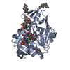 7ve0C 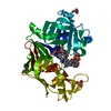 5yicS S: Starting model for refinement C: citing same article ( |
|---|---|
| Similar structure data |
-Links
 PDBj
PDBj- Assembly
| Deposited unit | 
| ||||||||
|---|---|---|---|---|---|---|---|---|---|
| 1 |
| ||||||||
| Unit cell |
|
-Components
| #1: Protein | Mass: 37113.926 Da / Num. of mol.: 1 Source method: isolated from a genetically manipulated source Source: (gene. exp.) Plasmodium falciparum (isolate 3D7) (eukaryote)Strain: isolate 3D7 / Gene: PF3D7_1408000 / Plasmid: pET32b / Production host:  Escherichia coli (E. coli) / Strain (production host): Rosetta-gami B(DE3)pLysS / References: UniProt: Q8I6V3, plasmepsin II Escherichia coli (E. coli) / Strain (production host): Rosetta-gami B(DE3)pLysS / References: UniProt: Q8I6V3, plasmepsin II | ||
|---|---|---|---|
| #2: Chemical | ChemComp-AB1 / Lopinavir  Mass: 628.801 Da / Num. of mol.: 1 / Source method: obtained synthetically / Formula: C37H48N4O5 / Feature type: SUBJECT OF INVESTIGATION / Comment: antiretroviral, protease inhibitor*YM Mass: 628.801 Da / Num. of mol.: 1 / Source method: obtained synthetically / Formula: C37H48N4O5 / Feature type: SUBJECT OF INVESTIGATION / Comment: antiretroviral, protease inhibitor*YM | ||
| #3: Chemical | CHAPS detergent  Mass: 614.877 Da / Num. of mol.: 2 / Source method: obtained synthetically / Formula: C32H58N2O7S / Comment: detergent*YM Mass: 614.877 Da / Num. of mol.: 2 / Source method: obtained synthetically / Formula: C32H58N2O7S / Comment: detergent*YMHas ligand of interest | Y | |
-Experimental details
-Experiment
| Experiment | Method: X-RAY DIFFRACTION / Number of used crystals: 1 |
|---|
- Sample preparation
| Crystal | Density Matthews: 2.81 Å3/Da / Density % sol: 56.33 % |
|---|---|
| Crystal grow | Temperature: 293 K / Method: vapor diffusion, sitting drop / pH: 5.5 / Details: 1.4 M ammonium sulfate, 0.1 M Bis-Tris pH 5.5 |
-Data collection
| Diffraction | Mean temperature: 100 K / Serial crystal experiment: N |
|---|---|
| Diffraction source | Source: ROTATING ANODE / Type: RIGAKU MICROMAX-007 HF / Wavelength: 1.5418 Å |
| Detector | Type: RIGAKU RAXIS IV++ / Detector: IMAGE PLATE / Date: Mar 21, 2020 |
| Radiation | Protocol: SINGLE WAVELENGTH / Monochromatic (M) / Laue (L): M / Scattering type: x-ray |
| Radiation wavelength | Wavelength: 1.5418 Å / Relative weight: 1 |
| Reflection | Resolution: 3.2→38.52 Å / Num. obs: 6877 / % possible obs: 99.9 % / Redundancy: 7.54 % / Biso Wilson estimate: 87.22 Å2 / CC1/2: 0.99 / Net I/σ(I): 9.95 |
| Reflection shell | Resolution: 3.2→3.3 Å / Redundancy: 7.57 % / Mean I/σ(I) obs: 1.04 / Num. unique obs: 602 / CC1/2: 0.37 / % possible all: 100 |
- Processing
| Software |
| ||||||||||||||||||||||||||||||||||||||||||||||||||||||||||||||||||||||||||||||||||||||||||||||||||||||||||||||||||||||||||||||||||||||||||||||||||||||||||||||||||||||||||||||||||||||
|---|---|---|---|---|---|---|---|---|---|---|---|---|---|---|---|---|---|---|---|---|---|---|---|---|---|---|---|---|---|---|---|---|---|---|---|---|---|---|---|---|---|---|---|---|---|---|---|---|---|---|---|---|---|---|---|---|---|---|---|---|---|---|---|---|---|---|---|---|---|---|---|---|---|---|---|---|---|---|---|---|---|---|---|---|---|---|---|---|---|---|---|---|---|---|---|---|---|---|---|---|---|---|---|---|---|---|---|---|---|---|---|---|---|---|---|---|---|---|---|---|---|---|---|---|---|---|---|---|---|---|---|---|---|---|---|---|---|---|---|---|---|---|---|---|---|---|---|---|---|---|---|---|---|---|---|---|---|---|---|---|---|---|---|---|---|---|---|---|---|---|---|---|---|---|---|---|---|---|---|---|---|---|---|
| Refinement | Method to determine structure: MOLECULAR REPLACEMENT Starting model: 5YIC Resolution: 3.2→38.52 Å / Cor.coef. Fo:Fc: 0.938 / Cor.coef. Fo:Fc free: 0.912 / SU B: 83.027 / SU ML: 0.637 / Cross valid method: THROUGHOUT / ESU R Free: 0.561 / Stereochemistry target values: MAXIMUM LIKELIHOOD / Details: HYDROGENS HAVE BEEN ADDED IN THE RIDING POSITIONS
| ||||||||||||||||||||||||||||||||||||||||||||||||||||||||||||||||||||||||||||||||||||||||||||||||||||||||||||||||||||||||||||||||||||||||||||||||||||||||||||||||||||||||||||||||||||||
| Solvent computation | Ion probe radii: 0.8 Å / Shrinkage radii: 0.8 Å / VDW probe radii: 1.2 Å / Solvent model: MASK | ||||||||||||||||||||||||||||||||||||||||||||||||||||||||||||||||||||||||||||||||||||||||||||||||||||||||||||||||||||||||||||||||||||||||||||||||||||||||||||||||||||||||||||||||||||||
| Displacement parameters | Biso mean: 127.423 Å2
| ||||||||||||||||||||||||||||||||||||||||||||||||||||||||||||||||||||||||||||||||||||||||||||||||||||||||||||||||||||||||||||||||||||||||||||||||||||||||||||||||||||||||||||||||||||||
| Refinement step | Cycle: 1 / Resolution: 3.2→38.52 Å
| ||||||||||||||||||||||||||||||||||||||||||||||||||||||||||||||||||||||||||||||||||||||||||||||||||||||||||||||||||||||||||||||||||||||||||||||||||||||||||||||||||||||||||||||||||||||
| Refine LS restraints |
|