 Movie
Movie Controller

[English] 日本語
 Yorodumi
- PDB-7uk1: Complex Structure of Human Polypyrimidine Splicing Factor (PSF/SF... -
+ Open data

- Basic information
| Entry | Database: PDB / ID: 7uk1 | ||||||
|---|---|---|---|---|---|---|---|
| Title | Complex Structure of Human Polypyrimidine Splicing Factor (PSF/SFPQ) with Murine Virus-like 30S Transcript-1 (VS30-1) Reveals Cooperative Binding of RNA | ||||||
 Components Components | Splicing factor, proline- and glutamine-rich | ||||||
 Keywords Keywords | SPLICING / Polypyridimine-binding splicing factor / 5'-polyuridine negative-sense (5'-PUN) template / RNA-dependent DNA-binding regulation / DNA-binding domain (DBD) / RNA-binding motif (RRM) / VL30-1 / Allostery / Cooperativity | ||||||
| Function / homology |  Function and homology information Function and homology informationPTK6 Regulates Proteins Involved in RNA Processing / negative regulation of circadian rhythm / alternative mRNA splicing, via spliceosome / Suppression of apoptosis / paraspeckles / positive regulation of oxidative stress-induced intrinsic apoptotic signaling pathway / activation of innate immune response / RNA splicing / double-strand break repair via homologous recombination / regulation of circadian rhythm ...PTK6 Regulates Proteins Involved in RNA Processing / negative regulation of circadian rhythm / alternative mRNA splicing, via spliceosome / Suppression of apoptosis / paraspeckles / positive regulation of oxidative stress-induced intrinsic apoptotic signaling pathway / activation of innate immune response / RNA splicing / double-strand break repair via homologous recombination / regulation of circadian rhythm / mRNA processing / nuclear matrix / histone deacetylase binding / RNA polymerase II transcription regulator complex / rhythmic process / transcription cis-regulatory region binding / nuclear speck / chromatin remodeling / innate immune response / negative regulation of DNA-templated transcription / chromatin binding / chromatin / regulation of DNA-templated transcription / negative regulation of transcription by RNA polymerase II / protein homodimerization activity / positive regulation of transcription by RNA polymerase II / DNA binding / RNA binding / nucleoplasm / nucleus / cytosolSimilarity search - Function | ||||||
| Biological species |  Homo sapiens (human) Homo sapiens (human) | ||||||
| Method | X-RAY DIFFRACTION / SYNCHROTRON / MOLECULAR REPLACEMENT / Resolution: 2.7 Å | ||||||
 Authors Authors | Lomakin, I.B. / Wang, J. | ||||||
| Funding support | 1items
| ||||||
 Citation Citation | Journal: Biochemistry / Year: 2022 Title: Insight into the Tumor Suppression Mechanism from the Structure of Human Polypyrimidine Splicing Factor (PSF/SFPQ) Complexed with a 30mer RNA from Murine Virus-like 30S Transcript-1. Authors: Wang, J. / Sachpatzidis, A. / Christian, T.D. / Lomakin, I.B. / Garen, A. / Konigsberg, W.H. | ||||||
| History |
|
- Structure visualization
| Structure viewer | Molecule: MolmilJmol/JSmol |
|---|
- Downloads & links
-Download
| PDBx/mmCIF format | 7uk1.cif.gz | 225.7 KB | Display | PDBx/mmCIF format |
|---|---|---|---|---|
| PDB format | pdb7uk1.ent.gz | 179.6 KB | Display | PDB format |
| PDBx/mmJSON format | 7uk1.json.gz | Tree view | PDBx/mmJSON format | |
| Others |  Other downloads Other downloads |
-Validation report
| Arichive directory | https://data.pdbj.org/pub/pdb/validation_reports/uk/7uk1ftp://data.pdbj.org/pub/pdb/validation_reports/uk/7uk1 | HTTPS FTP |
|---|
-Related structure data
| Related structure data | 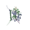 7uj1C  4wiiS S: Starting model for refinement C: citing same article ( |
|---|---|
| Similar structure data |
-Links
 PDBj
PDBj- Assembly
| Deposited unit | 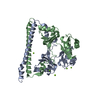
| ||||||||
|---|---|---|---|---|---|---|---|---|---|
| 1 |
| ||||||||
| Unit cell |
|
-Components
| #1: Protein | / 100 kDa DNA-pairing protein / hPOMp100 / DNA-binding p52/p100 complex / 100 kDa subunit / ...100 kDa DNA-pairing protein / hPOMp100 / DNA-binding p52/p100 complex / 100 kDa subunit / Polypyrimidine tract-binding protein-associated-splicing factor / PSF / PTB-associated-splicing factor Mass: 47764.684 Da / Num. of mol.: 2 Source method: isolated from a genetically manipulated source Source: (gene. exp.) Homo sapiens (human) / Gene: SFPQ, PSF / Production host:  Escherichia coli (E. coli) / References: UniProt: P23246 Escherichia coli (E. coli) / References: UniProt: P23246#2: Chemical | ChemComp-MG /   Mass: 24.305 Da / Num. of mol.: 5 / Source method: obtained synthetically / Formula: Mg Mass: 24.305 Da / Num. of mol.: 5 / Source method: obtained synthetically / Formula: Mg#3: Water | ChemComp-HOH / | Water Mass: 18.015 Da / Num. of mol.: 28 / Source method: isolated from a natural source / Formula: H2O Mass: 18.015 Da / Num. of mol.: 28 / Source method: isolated from a natural source / Formula: H2OHas ligand of interest | Y | |
|---|
-Experimental details
-Experiment
| Experiment | Method: X-RAY DIFFRACTION / Number of used crystals: 1 |
|---|
- Sample preparation
| Crystal grow | Temperature: 285.15 K / Method: evaporation / pH: 7 Details: 0.1 M MgCl2, 0.1 M HEPES pH 7.0, 15% (w/v),PEG4000, or 15% (w/v) PEG3500, or 20% PEG2000MME |
|---|
-Data collection
| Diffraction | Mean temperature: 80 K / Serial crystal experiment: N |
|---|---|
| Diffraction source | Source: SYNCHROTRON / Site: APS  / Beamline: 24-ID-C / Wavelength: 0.9792 Å / Beamline: 24-ID-C / Wavelength: 0.9792 Å |
| Detector | Type: DECTRIS PILATUS 6M / Detector: PIXEL / Date: Oct 15, 2016 |
| Radiation | Protocol: SINGLE WAVELENGTH / Monochromatic (M) / Laue (L): M / Scattering type: x-ray |
| Radiation wavelength | Wavelength: 0.9792 Å / Relative weight: 1 |
| Reflection | Resolution: 2.69→48.87 Å / Num. obs: 17804 / % possible obs: 98.9 % / Redundancy: 44.1 % / Rpim(I) all: 0.129 / Rrim(I) all: 0.91 / Net I/σ(I): 17.1 |
| Reflection shell | Resolution: 2.69→2.75 Å / Num. unique obs: 728 / CC1/2: 0.125 |
- Processing
| Software |
| |||||||||||||||||||||||||||||||||||||||||||||||||||||||||||||||||||||||||||||||||||||||||||||||||||||||||||||||||||||||||||||||||||||||||||||||||||||||||||||||||||||||||||||||
|---|---|---|---|---|---|---|---|---|---|---|---|---|---|---|---|---|---|---|---|---|---|---|---|---|---|---|---|---|---|---|---|---|---|---|---|---|---|---|---|---|---|---|---|---|---|---|---|---|---|---|---|---|---|---|---|---|---|---|---|---|---|---|---|---|---|---|---|---|---|---|---|---|---|---|---|---|---|---|---|---|---|---|---|---|---|---|---|---|---|---|---|---|---|---|---|---|---|---|---|---|---|---|---|---|---|---|---|---|---|---|---|---|---|---|---|---|---|---|---|---|---|---|---|---|---|---|---|---|---|---|---|---|---|---|---|---|---|---|---|---|---|---|---|---|---|---|---|---|---|---|---|---|---|---|---|---|---|---|---|---|---|---|---|---|---|---|---|---|---|---|---|---|---|---|---|---|
| Refinement | Method to determine structure: MOLECULAR REPLACEMENT Starting model: 4wii Resolution: 2.7→48.87 Å / Cor.coef. Fo:Fc: 0.967 / Cor.coef. Fo:Fc free: 0.909 / SU B: 54.257 / SU ML: 0.461 / Cross valid method: THROUGHOUT / σ(F): 0 / ESU R Free: 0.437 / Stereochemistry target values: MAXIMUM LIKELIHOOD / Details: U VALUES : WITH TLS ADDED
| |||||||||||||||||||||||||||||||||||||||||||||||||||||||||||||||||||||||||||||||||||||||||||||||||||||||||||||||||||||||||||||||||||||||||||||||||||||||||||||||||||||||||||||||
| Solvent computation | Ion probe radii: 0.8 Å / Shrinkage radii: 0.8 Å / VDW probe radii: 1.2 Å / Solvent model: MASK | |||||||||||||||||||||||||||||||||||||||||||||||||||||||||||||||||||||||||||||||||||||||||||||||||||||||||||||||||||||||||||||||||||||||||||||||||||||||||||||||||||||||||||||||
| Displacement parameters | Biso max: 342.66 Å2 / Biso mean: 112.829 Å2 / Biso min: 56.5 Å2
| |||||||||||||||||||||||||||||||||||||||||||||||||||||||||||||||||||||||||||||||||||||||||||||||||||||||||||||||||||||||||||||||||||||||||||||||||||||||||||||||||||||||||||||||
| Refinement step | Cycle: final / Resolution: 2.7→48.87 Å
| |||||||||||||||||||||||||||||||||||||||||||||||||||||||||||||||||||||||||||||||||||||||||||||||||||||||||||||||||||||||||||||||||||||||||||||||||||||||||||||||||||||||||||||||
| Refine LS restraints |
| |||||||||||||||||||||||||||||||||||||||||||||||||||||||||||||||||||||||||||||||||||||||||||||||||||||||||||||||||||||||||||||||||||||||||||||||||||||||||||||||||||||||||||||||
| LS refinement shell | Resolution: 2.7→2.77 Å / Rfactor Rfree error: 0 / Total num. of bins used: 20
| |||||||||||||||||||||||||||||||||||||||||||||||||||||||||||||||||||||||||||||||||||||||||||||||||||||||||||||||||||||||||||||||||||||||||||||||||||||||||||||||||||||||||||||||
| Refinement TLS params. | Method: refined / Refine-ID: X-RAY DIFFRACTION
| |||||||||||||||||||||||||||||||||||||||||||||||||||||||||||||||||||||||||||||||||||||||||||||||||||||||||||||||||||||||||||||||||||||||||||||||||||||||||||||||||||||||||||||||
| Refinement TLS group |
|
