 Movie
Movie Controller


+ Open data

- Basic information
| Entry | Database: PDB / ID: 7ujd | ||||||
|---|---|---|---|---|---|---|---|
| Title | PSMD2 Structure bound to MC1 and Fab8/14 | ||||||
 Components Components |
| ||||||
 Keywords Keywords | PROTEIN BINDING/Immune System / 26S proteasome macrocycle /  PROTEIN BINDING / PROTEIN BINDING-Immune System complex PROTEIN BINDING / PROTEIN BINDING-Immune System complex | ||||||
| Function / homology |  Function and homology information Function and homology informationproteasome accessory complex / proteasome regulatory particle / proteasome regulatory particle, base subcomplex / Regulation of ornithine decarboxylase (ODC) / Cross-presentation of soluble exogenous antigens (endosomes) / Somitogenesis / regulation of protein catabolic process / proteasome storage granule / enzyme regulator activity / proteasome complex ...proteasome accessory complex / proteasome regulatory particle / proteasome regulatory particle, base subcomplex / Regulation of ornithine decarboxylase (ODC) / Cross-presentation of soluble exogenous antigens (endosomes) / Somitogenesis / regulation of protein catabolic process / proteasome storage granule / enzyme regulator activity / proteasome complex / Regulation of activated PAK-2p34 by proteasome mediated degradation / Autodegradation of Cdh1 by Cdh1:APC/C / APC/C:Cdc20 mediated degradation of Securin / Asymmetric localization of PCP proteins / SCF-beta-TrCP mediated degradation of Emi1 / NIK-->noncanonical NF-kB signaling / Ubiquitin-dependent degradation of Cyclin D / AUF1 (hnRNP D0) binds and destabilizes mRNA / TNFR2 non-canonical NF-kB pathway / Assembly of the pre-replicative complex / Vpu mediated degradation of CD4 / Degradation of DVL / Ubiquitin Mediated Degradation of Phosphorylated Cdc25A / Dectin-1 mediated noncanonical NF-kB signaling / Hh mutants are degraded by ERAD / Cdc20:Phospho-APC/C mediated degradation of Cyclin A / Degradation of AXIN / Defective CFTR causes cystic fibrosis / Degradation of GLI1 by the proteasome / Activation of NF-kappaB in B cells / Hedgehog ligand biogenesis / Negative regulation of NOTCH4 signaling / G2/M Checkpoints / GSK3B and BTRC:CUL1-mediated-degradation of NFE2L2 / Autodegradation of the E3 ubiquitin ligase COP1 / Vif-mediated degradation of APOBEC3G / Hedgehog 'on' state / Regulation of RUNX3 expression and activity / Degradation of GLI2 by the proteasome / GLI3 is processed to GLI3R by the proteasome / MAPK6/MAPK4 signaling / FBXL7 down-regulates AURKA during mitotic entry and in early mitosis / APC/C:Cdh1 mediated degradation of Cdc20 and other APC/C:Cdh1 targeted proteins in late mitosis/early G1 / ABC-family proteins mediated transport / Degradation of beta-catenin by the destruction complex / Oxygen-dependent proline hydroxylation of Hypoxia-inducible Factor Alpha / CDK-mediated phosphorylation and removal of Cdc6 / CLEC7A (Dectin-1) signaling / SCF(Skp2)-mediated degradation of p27/p21 / Regulation of expression of SLITs and ROBOs / FCERI mediated NF-kB activation / Regulation of PTEN stability and activity / Interleukin-1 signaling / Orc1 removal from chromatin / Regulation of RAS by GAPs / Separation of Sister Chromatids / Regulation of RUNX2 expression and activity / UCH proteinases / The role of GTSE1 in G2/M progression after G2 checkpoint / KEAP1-NFE2L2 pathway / Antigen processing: Ubiquitination & Proteasome degradation / Downstream TCR signaling / Neddylation / RUNX1 regulates transcription of genes involved in differentiation of HSCs / ER-Phagosome pathway / proteasome-mediated ubiquitin-dependent protein catabolic process / secretory granule lumen / ficolin-1-rich granule lumen / Ub-specific processing proteases / Neutrophil degranulation / extracellular exosome / extracellular region / nucleoplasm / membrane / nucleus / cytosolSimilarity search - Function | ||||||
| Biological species |  Homo sapiens (human) Homo sapiens (human)synthetic construct (others) | ||||||
| Method | ELECTRON MICROSCOPY / single particle reconstruction / cryo EM / Resolution: 2.5 Å | ||||||
 Authors Authors | Johnson, M.C. / Bashore, C. / Ciferri, C. / Dueber, E.C. | ||||||
| Funding support | 1items
| ||||||
 Citation Citation | Journal: Nat Chem Biol / Year: 2023 Title: Targeted degradation via direct 26S proteasome recruitment. Authors: Charlene Bashore / Sumit Prakash / Matthew C Johnson / Ryan J Conrad / Ivy A Kekessie / Suzie J Scales / Noriko Ishisoko / Tracy Kleinheinz / Peter S Liu / Nataliya Popovych / Aaron T ...Authors: Charlene Bashore / Sumit Prakash / Matthew C Johnson / Ryan J Conrad / Ivy A Kekessie / Suzie J Scales / Noriko Ishisoko / Tracy Kleinheinz / Peter S Liu / Nataliya Popovych / Aaron T Wecksler / Lijuan Zhou / Christine Tam / Inna Zilberleyb / Rajini Srinivasan / Robert A Blake / Aimin Song / Steven T Staben / Yingnan Zhang / David Arnott / Wayne J Fairbrother / Scott A Foster / Ingrid E Wertz / Claudio Ciferri / Erin C Dueber /  Abstract: Engineered destruction of target proteins by recruitment to the cell's degradation machinery has emerged as a promising strategy in drug discovery. The majority of molecules that facilitate targeted ...Engineered destruction of target proteins by recruitment to the cell's degradation machinery has emerged as a promising strategy in drug discovery. The majority of molecules that facilitate targeted degradation do so via a select number of ubiquitin ligases, restricting this therapeutic approach to tissue types that express the requisite ligase. Here, we describe a new strategy of targeted protein degradation through direct substrate recruitment to the 26S proteasome. The proteolytic complex is essential and abundantly expressed in all cells; however, proteasomal ligands remain scarce. We identify potent peptidic macrocycles that bind directly to the 26S proteasome subunit PSMD2, with a 2.5-Å-resolution cryo-electron microscopy complex structure revealing a binding site near the 26S pore. Conjugation of this macrocycle to a potent BRD4 ligand enabled generation of chimeric molecules that effectively degrade BRD4 in cells, thus demonstrating that degradation via direct proteasomal recruitment is a viable strategy for targeted protein degradation. | ||||||
| History |
|
- Structure visualization
| Structure viewer | Molecule: MolmilJmol/JSmol |
|---|
- Downloads & links
-Download
| PDBx/mmCIF format | 7ujd.cif.gz | 139.9 KB | Display | PDBx/mmCIF format |
|---|---|---|---|---|
| PDB format | pdb7ujd.ent.gz | 101.9 KB | Display | PDB format |
| PDBx/mmJSON format | 7ujd.json.gz | Tree view | PDBx/mmJSON format | |
| Others |  Other downloads Other downloads |
-Validation report
| Arichive directory | https://data.pdbj.org/pub/pdb/validation_reports/uj/7ujdftp://data.pdbj.org/pub/pdb/validation_reports/uj/7ujd | HTTPS FTP |
|---|
-Related structure data
| Related structure data |  24742MC 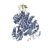 7uihC C: citing same article ( M: map data used to model this data |
|---|---|
| Similar structure data |
-Links
 PDBj
PDBj


- Assembly
| Deposited unit | 
|
|---|---|
| 1 |
|
-Components
-Antibody , 4 types, 4 molecules CDEF
| #2: Antibody | Mass: 23825.389 Da / Num. of mol.: 1 Source method: isolated from a genetically manipulated source Source: (gene. exp.) Homo sapiens (human) / Production host:  Escherichia coli (E. coli) Escherichia coli (E. coli) |
|---|---|
| #3: Antibody | Mass: 24717.482 Da / Num. of mol.: 1 Source method: isolated from a genetically manipulated source Source: (gene. exp.) Homo sapiens (human) / Production host: Escherichia coli (E. coli) |
| #4: Antibody | Mass: 23689.238 Da / Num. of mol.: 1 Source method: isolated from a genetically manipulated source Source: (gene. exp.) Homo sapiens (human) / Production host: Escherichia coli (E. coli) |
| #5: Antibody | Mass: 24606.496 Da / Num. of mol.: 1 Source method: isolated from a genetically manipulated source Source: (gene. exp.) Homo sapiens (human) / Production host: Escherichia coli (E. coli) |
-Protein / Protein/peptide , 2 types, 2 molecules AZ
| #1: Protein | Mass: 70199.539 Da / Num. of mol.: 1 Source method: isolated from a genetically manipulated source Source: (gene. exp.) Homo sapiens (human) / Gene: PSMD2, TRAP2 / Production host: Escherichia coli (E. coli) / References: UniProt: Q13200 |
|---|---|
| #6: Protein/peptide | Mass: 1864.071 Da / Num. of mol.: 1 / Source method: obtained synthetically / Source: (synth.) synthetic construct (others) |
-Details
| Has ligand of interest | Y |
|---|
-Experimental details
-Experiment
| Experiment | Method: ELECTRON MICROSCOPY |
|---|---|
| EM experiment | Aggregation state: PARTICLE / 3D reconstruction method: single particle reconstruction |
- Sample preparation
| Component | Name: PSMD2 bound to MC1 and Fab8/14 / Type: ORGANELLE OR CELLULAR COMPONENT / Entity ID: all / Source: RECOMBINANT |
|---|---|
| Source (natural) | Organism: Homo sapiens (human) |
| Source (recombinant) | Organism: Escherichia coli (E. coli) |
| Buffer solution | pH: 7.5 |
| Specimen | Embedding applied: NO / Shadowing applied: NO / Staining applied: NO / Vitrification applied: YES |
| Vitrification | Cryogen name: ETHANE |
- Electron microscopy imaging
| Experimental equipment |  Model: Titan Krios / Image courtesy: FEI Company |
|---|---|
| Microscopy | Model: FEI TITAN KRIOS |
| Electron gun | Electron source: FIELD EMISSION GUN / Accelerating voltage: 300 kV / Illumination mode: OTHER |
| Electron lens | Mode: OTHER / Nominal defocus max: 3000 nm / Nominal defocus min: 300 nm |
| Image recording | Electron dose: 64 e/Å2 / Film or detector model: GATAN K3 BIOQUANTUM (6k x 4k) |
- Processing
| CTF correction | Type: PHASE FLIPPING AND AMPLITUDE CORRECTION |
|---|---|
| 3D reconstruction | Resolution: 2.5 Å / Resolution method: FSC 0.143 CUT-OFF / Num. of particles: 105705 / Symmetry type: POINT |
