 Movie
Movie Controller

[English] 日本語
 Yorodumi
- PDB-7uf3: RibB from Vibrio cholera bound with L-xylulose-5-phosphate (L-Xy5... -
+ Open data

- Basic information
| Entry | Database: PDB / ID: 7uf3 | ||||||
|---|---|---|---|---|---|---|---|
| Title | RibB from Vibrio cholera bound with L-xylulose-5-phosphate (L-Xy5P) and manganese | ||||||
 Components Components | 3,4-dihydroxy-2-butanone 4-phosphate synthase | ||||||
 Keywords Keywords |  LYASE / RibB / riboflavin / 3 / 4-dihydroxy-2-butanone 4-phosphate synthase LYASE / RibB / riboflavin / 3 / 4-dihydroxy-2-butanone 4-phosphate synthase | ||||||
| Function / homology |  Function and homology information3,4-dihydroxy-2-butanone-4-phosphate synthase / 3,4-dihydroxy-2-butanone-4-phosphate synthase activity / riboflavin biosynthetic process / manganese ion binding / magnesium ion binding Function and homology information3,4-dihydroxy-2-butanone-4-phosphate synthase / 3,4-dihydroxy-2-butanone-4-phosphate synthase activity / riboflavin biosynthetic process / manganese ion binding / magnesium ion bindingSimilarity search - Function | ||||||
| Biological species |   Vibrio cholerae (bacteria) Vibrio cholerae (bacteria) | ||||||
| Method | X-RAY DIFFRACTION / SYNCHROTRON / MOLECULAR REPLACEMENT / Resolution: 2 Å | ||||||
 Authors Authors | Kenjic, N. / Meneely, K.M. / Lamb, A.L. | ||||||
| Funding support |  United States, 1items United States, 1items
| ||||||
 Citation Citation | Journal: J.Am.Chem.Soc. / Year: 2022 Title: Evidence for the Chemical Mechanism of RibB (3,4-Dihydroxy-2-butanone 4-phosphate Synthase) of Riboflavin Biosynthesis. Authors: Kenjic, N. / Meneely, K.M. / Wherritt, D.J. / Denler, M.C. / Jackson, T.A. / Moran, G.R. / Lamb, A.L. | ||||||
| History |
|
- Structure visualization
| Structure viewer | Molecule: MolmilJmol/JSmol |
|---|
- Downloads & links
-Download
| PDBx/mmCIF format | 7uf3.cif.gz | 96 KB | Display | PDBx/mmCIF format |
|---|---|---|---|---|
| PDB format | pdb7uf3.ent.gz | 70.8 KB | Display | PDB format |
| PDBx/mmJSON format | 7uf3.json.gz | Tree view | PDBx/mmJSON format | |
| Others |  Other downloads Other downloads |
-Validation report
| Arichive directory | https://data.pdbj.org/pub/pdb/validation_reports/uf/7uf3ftp://data.pdbj.org/pub/pdb/validation_reports/uf/7uf3 | HTTPS FTP |
|---|
-Related structure data
| Related structure data | 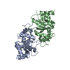 7uezC 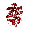 7uf0C 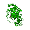 7uf1C  7uf2C 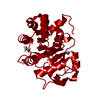 7uf4C  7uf5C 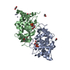 4p8eS S: Starting model for refinement C: citing same article ( |
|---|---|
| Similar structure data |
-Links
 PDBj
PDBj- Assembly
| Deposited unit | 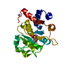
| ||||||||||||
|---|---|---|---|---|---|---|---|---|---|---|---|---|---|
| 1 | 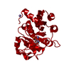
| ||||||||||||
| Unit cell |
| ||||||||||||
| Components on special symmetry positions |
|
-Components
| #1: Protein | Mass: 23589.953 Da / Num. of mol.: 1 Source method: isolated from a genetically manipulated source Source: (gene. exp.) Vibrio cholerae (bacteria)Gene: ribB, D6U24_02240, ERS013198_00268, ERS013199_01247, ERS013202_00910, ERS013207_00669, FXE67_08125 Production host: Escherichia coli (E. coli)References: UniProt: A0A0H6NPW4, 3,4-dihydroxy-2-butanone-4-phosphate synthase | ||||
|---|---|---|---|---|---|
| #2: Sugar | ChemComp-N3U / 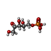  Type: L-saccharide / Mass: 230.110 Da / Num. of mol.: 1 / Source method: obtained synthetically / Formula: C5H11O8P / Feature type: SUBJECT OF INVESTIGATION Type: L-saccharide / Mass: 230.110 Da / Num. of mol.: 1 / Source method: obtained synthetically / Formula: C5H11O8P / Feature type: SUBJECT OF INVESTIGATION | ||||
| #3: Chemical |   Mass: 54.938 Da / Num. of mol.: 3 / Source method: obtained synthetically / Formula: Mn Mass: 54.938 Da / Num. of mol.: 3 / Source method: obtained synthetically / Formula: Mn#4: Water | ChemComp-HOH / | Water Mass: 18.015 Da / Num. of mol.: 122 / Source method: isolated from a natural source / Formula: H2O Mass: 18.015 Da / Num. of mol.: 122 / Source method: isolated from a natural source / Formula: H2OHas ligand of interest | Y | |
-Experimental details
-Experiment
| Experiment | Method: X-RAY DIFFRACTION / Number of used crystals: 1 |
|---|
- Sample preparation
| Crystal | Density Matthews: 2.31 Å3/Da / Density % sol: 46.73 % |
|---|---|
| Crystal grow | Temperature: 298 K / Method: vapor diffusion, hanging drop / Details: 0.1 M lithium acetate, 12-18% w/v PEG3350 |
-Data collection
| Diffraction | Mean temperature: 100 K / Serial crystal experiment: N |
|---|---|
| Diffraction source | Source: SYNCHROTRON / Site: SSRL / Beamline: BL9-2 / Wavelength: 0.97946 Å |
| Detector | Type: DECTRIS PILATUS 6M / Detector: PIXEL / Date: Feb 8, 2019 |
| Radiation | Protocol: SINGLE WAVELENGTH / Monochromatic (M) / Laue (L): M / Scattering type: x-ray |
| Radiation wavelength | Wavelength: 0.97946 Å / Relative weight: 1 |
| Reflection | Resolution: 2→39.51 Å / Num. obs: 15092 / % possible obs: 99.6 % / Redundancy: 6.6 % / Biso Wilson estimate: 23.26 Å2 / Rpim(I) all: 0.043 / Net I/σ(I): 16.9 |
| Reflection shell | Resolution: 2→2.05 Å / Mean I/σ(I) obs: 3.8 / Num. unique obs: 1060 / Rpim(I) all: 0.256 |
- Processing
| Software |
| ||||||||||||||||||||||||||||||||||||||||||||||||||||||||||||||||||||||||||||||||||||
|---|---|---|---|---|---|---|---|---|---|---|---|---|---|---|---|---|---|---|---|---|---|---|---|---|---|---|---|---|---|---|---|---|---|---|---|---|---|---|---|---|---|---|---|---|---|---|---|---|---|---|---|---|---|---|---|---|---|---|---|---|---|---|---|---|---|---|---|---|---|---|---|---|---|---|---|---|---|---|---|---|---|---|---|---|---|
| Refinement | Method to determine structure: MOLECULAR REPLACEMENT Starting model: PDB entry 4P8E Resolution: 2→39.51 Å / SU ML: 0.1852 / Cross valid method: FREE R-VALUE / σ(F): 1.33 / Phase error: 20.1933 Stereochemistry target values: GeoStd + Monomer Library + CDL v1.2
| ||||||||||||||||||||||||||||||||||||||||||||||||||||||||||||||||||||||||||||||||||||
| Solvent computation | Shrinkage radii: 0.9 Å / VDW probe radii: 1.1 Å / Solvent model: FLAT BULK SOLVENT MODEL | ||||||||||||||||||||||||||||||||||||||||||||||||||||||||||||||||||||||||||||||||||||
| Displacement parameters | Biso mean: 26.69 Å2 | ||||||||||||||||||||||||||||||||||||||||||||||||||||||||||||||||||||||||||||||||||||
| Refinement step | Cycle: LAST / Resolution: 2→39.51 Å
| ||||||||||||||||||||||||||||||||||||||||||||||||||||||||||||||||||||||||||||||||||||
| Refine LS restraints |
| ||||||||||||||||||||||||||||||||||||||||||||||||||||||||||||||||||||||||||||||||||||
| LS refinement shell |
|
