 Movie
Movie Controller

[English] 日本語
 Yorodumi
- PDB-7tyg: Structure of the human leucine rich repeat protein SHOC2, residue... -
+ Open data

- Basic information
| Entry | Database: PDB / ID: 7tyg | ||||||
|---|---|---|---|---|---|---|---|
| Title | Structure of the human leucine rich repeat protein SHOC2, residues 80-582 | ||||||
 Components Components | Leucine-rich repeat protein SHOC-2 | ||||||
 Keywords Keywords | HYDROLASE / RAS / PP1 / SHOC2 / LRR | ||||||
| Function / homology |  Function and homology information Function and homology informationcellular response to growth hormone stimulus / protein phosphatase type 1 complex / negative regulation of neural precursor cell proliferation / nerve growth factor signaling pathway / protein phosphatase 1 binding / protein phosphatase regulator activity / SHOC2 M1731 mutant abolishes MRAS complex function / Gain-of-function MRAS complexes activate RAF signaling / positive regulation of Ras protein signal transduction / negative regulation of neuron differentiation ...cellular response to growth hormone stimulus / protein phosphatase type 1 complex / negative regulation of neural precursor cell proliferation / nerve growth factor signaling pathway / protein phosphatase 1 binding / protein phosphatase regulator activity / SHOC2 M1731 mutant abolishes MRAS complex function / Gain-of-function MRAS complexes activate RAF signaling / positive regulation of Ras protein signal transduction / negative regulation of neuron differentiation / fibroblast growth factor receptor signaling pathway / positive regulation of neuron differentiation / RAF activation / positive regulation of neuron projection development / protein phosphatase binding / Ras protein signal transduction / signal transduction / nucleoplasm / nucleus / cytosol / cytoplasmSimilarity search - Function | ||||||
| Biological species |  Homo sapiens (human) Homo sapiens (human) | ||||||
| Method | X-RAY DIFFRACTION / SYNCHROTRON / MOLECULAR REPLACEMENT / Resolution: 1.9 Å | ||||||
 Authors Authors | Dhembi, A. / Clark, K. / King, D.A. | ||||||
| Funding support | 1items
| ||||||
 Citation Citation | Journal: Nature / Year: 2022 Title: Structure of the MRAS-SHOC2-PP1C phosphatase complex. Authors: Hauseman, Z.J. / Fodor, M. / Dhembi, A. / Viscomi, J. / Egli, D. / Bleu, M. / Katz, S. / Park, E. / Jang, D.M. / Porter, K.A. / Meili, F. / Guo, H. / Kerr, G. / Molle, S. / Velez-Vega, C. / ...Authors: Hauseman, Z.J. / Fodor, M. / Dhembi, A. / Viscomi, J. / Egli, D. / Bleu, M. / Katz, S. / Park, E. / Jang, D.M. / Porter, K.A. / Meili, F. / Guo, H. / Kerr, G. / Molle, S. / Velez-Vega, C. / Beyer, K.S. / Galli, G.G. / Maira, S.M. / Stams, T. / Clark, K. / Eck, M.J. / Tordella, L. / Thoma, C.R. / King, D.A. | ||||||
| History |
|
- Structure visualization
| Structure viewer | Molecule: MolmilJmol/JSmol |
|---|
- Downloads & links
-Download
| PDBx/mmCIF format | 7tyg.cif.gz | 500.7 KB | Display | PDBx/mmCIF format |
|---|---|---|---|---|
| PDB format | pdb7tyg.ent.gz | 338.8 KB | Display | PDB format |
| PDBx/mmJSON format | 7tyg.json.gz | Tree view | PDBx/mmJSON format | |
| Others |  Other downloads Other downloads |
-Validation report
| Arichive directory | https://data.pdbj.org/pub/pdb/validation_reports/ty/7tygftp://data.pdbj.org/pub/pdb/validation_reports/ty/7tyg | HTTPS FTP |
|---|
-Related structure data
| Related structure data | 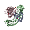 7txhC  4tzhS S: Starting model for refinement C: citing same article ( |
|---|---|
| Similar structure data |
-Links
 PDBj
PDBj


- Assembly
| Deposited unit | 
| ||||||||||||
|---|---|---|---|---|---|---|---|---|---|---|---|---|---|
| 1 | 
| ||||||||||||
| 2 | 
| ||||||||||||
| Unit cell |
|
-Components
| #1: Protein | / Protein soc-2 homolog / Protein sur-8 homolog Mass: 56737.582 Da / Num. of mol.: 2 / Fragment: residues 80-582 Source method: isolated from a genetically manipulated source Source: (gene. exp.) Homo sapiens (human) / Gene: SHOC2, KIAA0862 / Production host:   Spodoptera frugiperda (fall armyworm) / References: UniProt: Q9UQ13 Spodoptera frugiperda (fall armyworm) / References: UniProt: Q9UQ13#2: Chemical | ChemComp-MG / |   Mass: 24.305 Da / Num. of mol.: 1 / Source method: isolated from a natural source / Formula: Mg Mass: 24.305 Da / Num. of mol.: 1 / Source method: isolated from a natural source / Formula: Mg#3: Water | ChemComp-HOH / | Water Mass: 18.015 Da / Num. of mol.: 625 / Source method: isolated from a natural source / Formula: H2O Mass: 18.015 Da / Num. of mol.: 625 / Source method: isolated from a natural source / Formula: H2OHas ligand of interest | N | |
|---|
-Experimental details
-Experiment
| Experiment | Method: X-RAY DIFFRACTION / Number of used crystals: 1 |
|---|
- Sample preparation
| Crystal | Density Matthews: 2.45 Å3/Da / Density % sol: 49.87 % |
|---|---|
| Crystal grow | Temperature: 294 K / Method: vapor diffusion, sitting drop / pH: 8 Details: 0.1M Tris-Cl(8.0), 0.2 M MgCl2, 11% PEG4K, 2.5% 1,5-diaminopentane |
-Data collection
| Diffraction | Mean temperature: 100 K / Serial crystal experiment: N |
|---|---|
| Diffraction source | Source: SYNCHROTRON / Site: APS  / Beamline: 17-ID / Wavelength: 1 Å / Beamline: 17-ID / Wavelength: 1 Å |
| Detector | Type: DECTRIS EIGER X 9M / Detector: PIXEL / Date: Dec 20, 2020 |
| Radiation | Protocol: SINGLE WAVELENGTH / Monochromatic (M) / Laue (L): M / Scattering type: x-ray |
| Radiation wavelength | Wavelength: 1 Å / Relative weight: 1 |
| Reflection | Resolution: 1.895→118.303 Å / Num. obs: 82107 / % possible obs: 94.7 % / Redundancy: 3.3 % / Biso Wilson estimate: 34.29 Å2 / CC1/2: 0.998 / Rmerge(I) obs: 0.051 / Rrim(I) all: 0.061 / Net I/σ(I): 13 |
| Reflection shell | Resolution: 1.895→1.902 Å / Redundancy: 3.3 % / Rmerge(I) obs: 0.539 / Mean I/σ(I) obs: 2 / Num. unique obs: 869 / CC1/2: 0.748 / Rrim(I) all: 0.644 / % possible all: 100 |
- Processing
| Software |
| ||||||||||||||||||||||||||||||||||||||||||||||||||||||||||||||||||||||||||||||||||||||||||||||||||||||||||||||||||||||||||||||||||||||||||||||||||||||||||||||||||||||||||||||||||||||||||||||||||||||||||||||||||
|---|---|---|---|---|---|---|---|---|---|---|---|---|---|---|---|---|---|---|---|---|---|---|---|---|---|---|---|---|---|---|---|---|---|---|---|---|---|---|---|---|---|---|---|---|---|---|---|---|---|---|---|---|---|---|---|---|---|---|---|---|---|---|---|---|---|---|---|---|---|---|---|---|---|---|---|---|---|---|---|---|---|---|---|---|---|---|---|---|---|---|---|---|---|---|---|---|---|---|---|---|---|---|---|---|---|---|---|---|---|---|---|---|---|---|---|---|---|---|---|---|---|---|---|---|---|---|---|---|---|---|---|---|---|---|---|---|---|---|---|---|---|---|---|---|---|---|---|---|---|---|---|---|---|---|---|---|---|---|---|---|---|---|---|---|---|---|---|---|---|---|---|---|---|---|---|---|---|---|---|---|---|---|---|---|---|---|---|---|---|---|---|---|---|---|---|---|---|---|---|---|---|---|---|---|---|---|---|---|---|---|---|
| Refinement | Method to determine structure: MOLECULAR REPLACEMENT Starting model: 4TZH Resolution: 1.9→41.44 Å / SU ML: 0.2322 / Cross valid method: FREE R-VALUE / σ(F): 1.36 / Phase error: 24.4087 Stereochemistry target values: GeoStd + Monomer Library + CDL v1.2
| ||||||||||||||||||||||||||||||||||||||||||||||||||||||||||||||||||||||||||||||||||||||||||||||||||||||||||||||||||||||||||||||||||||||||||||||||||||||||||||||||||||||||||||||||||||||||||||||||||||||||||||||||||
| Solvent computation | Shrinkage radii: 0.9 Å / VDW probe radii: 1.11 Å / Solvent model: FLAT BULK SOLVENT MODEL | ||||||||||||||||||||||||||||||||||||||||||||||||||||||||||||||||||||||||||||||||||||||||||||||||||||||||||||||||||||||||||||||||||||||||||||||||||||||||||||||||||||||||||||||||||||||||||||||||||||||||||||||||||
| Displacement parameters | Biso mean: 40.44 Å2 | ||||||||||||||||||||||||||||||||||||||||||||||||||||||||||||||||||||||||||||||||||||||||||||||||||||||||||||||||||||||||||||||||||||||||||||||||||||||||||||||||||||||||||||||||||||||||||||||||||||||||||||||||||
| Refinement step | Cycle: LAST / Resolution: 1.9→41.44 Å
| ||||||||||||||||||||||||||||||||||||||||||||||||||||||||||||||||||||||||||||||||||||||||||||||||||||||||||||||||||||||||||||||||||||||||||||||||||||||||||||||||||||||||||||||||||||||||||||||||||||||||||||||||||
| Refine LS restraints |
| ||||||||||||||||||||||||||||||||||||||||||||||||||||||||||||||||||||||||||||||||||||||||||||||||||||||||||||||||||||||||||||||||||||||||||||||||||||||||||||||||||||||||||||||||||||||||||||||||||||||||||||||||||
| LS refinement shell |
| ||||||||||||||||||||||||||||||||||||||||||||||||||||||||||||||||||||||||||||||||||||||||||||||||||||||||||||||||||||||||||||||||||||||||||||||||||||||||||||||||||||||||||||||||||||||||||||||||||||||||||||||||||
| Refinement TLS params. | Method: refined / Origin x: -31.3239005291 Å / Origin y: 1.33645829092 Å / Origin z: 28.8237093738 Å
| ||||||||||||||||||||||||||||||||||||||||||||||||||||||||||||||||||||||||||||||||||||||||||||||||||||||||||||||||||||||||||||||||||||||||||||||||||||||||||||||||||||||||||||||||||||||||||||||||||||||||||||||||||
| Refinement TLS group | Selection details: all |
