 Movie
Movie Controller

[English] 日本語
 Yorodumi
- PDB-7tuo: Crystal structure analysis of human USP28 complex with a compound -
+ Open data

- Basic information
| Entry | Database: PDB / ID: 7tuo | ||||||
|---|---|---|---|---|---|---|---|
| Title | Crystal structure analysis of human USP28 complex with a compound | ||||||
 Components Components | Ubiquitin carboxyl-terminal hydrolase 28 | ||||||
 Keywords Keywords | HYDROLASE/HYDROLASE INHIBITOR /  deubiquitinase / inhibitor / protein-inhibitor complex / HYDROLASE-HYDROLASE INHIBITOR complex deubiquitinase / inhibitor / protein-inhibitor complex / HYDROLASE-HYDROLASE INHIBITOR complex | ||||||
| Function / homology |  Function and homology information Function and homology informationprotein deubiquitination involved in ubiquitin-dependent protein catabolic process / deubiquitinase activity / response to ionizing radiation / protein deubiquitination / intrinsic apoptotic signaling pathway in response to DNA damage by p53 class mediator / DNA damage checkpoint signaling / regulation of protein stability / cellular response to UV / ubiquitinyl hydrolase 1 / cell population proliferation ...protein deubiquitination involved in ubiquitin-dependent protein catabolic process / deubiquitinase activity / response to ionizing radiation / protein deubiquitination / intrinsic apoptotic signaling pathway in response to DNA damage by p53 class mediator / DNA damage checkpoint signaling / regulation of protein stability / cellular response to UV / ubiquitinyl hydrolase 1 / cell population proliferation / cysteine-type deubiquitinase activity / nuclear body / Ub-specific processing proteases / DNA repair / DNA damage response / protein-containing complex / nucleoplasm / nucleus / cytosolSimilarity search - Function | ||||||
| Biological species |  Homo sapiens (human) Homo sapiens (human) | ||||||
| Method | X-RAY DIFFRACTION / SYNCHROTRON / MOLECULAR REPLACEMENT / molecular replacement / Resolution: 1.96 Å | ||||||
 Authors Authors | Seo, H.-S. / Dhe-Paganon, S. | ||||||
| Funding support |  United States, 1items United States, 1items
| ||||||
 Citation Citation | Journal: To Be Published Title: Crystal Structure Analysis of human USP28 complex with a compound Authors: Seo, H.-S. / Dhe-Paganon, S. | ||||||
| History |
|
- Structure visualization
| Structure viewer | Molecule: MolmilJmol/JSmol |
|---|
- Downloads & links
-Download
| PDBx/mmCIF format | 7tuo.cif.gz | 159.1 KB | Display | PDBx/mmCIF format |
|---|---|---|---|---|
| PDB format | pdb7tuo.ent.gz | 122.2 KB | Display | PDB format |
| PDBx/mmJSON format | 7tuo.json.gz | Tree view | PDBx/mmJSON format | |
| Others |  Other downloads Other downloads |
-Validation report
| Arichive directory | https://data.pdbj.org/pub/pdb/validation_reports/tu/7tuoftp://data.pdbj.org/pub/pdb/validation_reports/tu/7tuo | HTTPS FTP |
|---|
-Related structure data
| Related structure data | 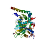 6hehS S: Starting model for refinement |
|---|---|
| Similar structure data |
-Links
 PDBj
PDBj

- Assembly
| Deposited unit | 
| ||||||||
|---|---|---|---|---|---|---|---|---|---|
| 1 |
| ||||||||
| Unit cell |
| ||||||||
| Components on special symmetry positions |
|
-Components
| #1: Protein | Mass: 44972.109 Da / Num. of mol.: 1 Source method: isolated from a genetically manipulated source Source: (gene. exp.) Homo sapiens (human) / Gene: USP28, KIAA1515 / Production host:  Escherichia coli BL21(DE3) (bacteria) / References: UniProt: Q96RU2, ubiquitinyl hydrolase 1 Escherichia coli BL21(DE3) (bacteria) / References: UniProt: Q96RU2, ubiquitinyl hydrolase 1 | ||||||
|---|---|---|---|---|---|---|---|
| #2: Chemical |   Mass: 421.558 Da / Num. of mol.: 2 / Source method: obtained synthetically / Formula: C23H27N5OS / Feature type: SUBJECT OF INVESTIGATION Mass: 421.558 Da / Num. of mol.: 2 / Source method: obtained synthetically / Formula: C23H27N5OS / Feature type: SUBJECT OF INVESTIGATION#3: Chemical | ChemComp-CL / | Chloride  Mass: 35.453 Da / Num. of mol.: 1 / Source method: obtained synthetically / Formula: Cl Mass: 35.453 Da / Num. of mol.: 1 / Source method: obtained synthetically / Formula: Cl#4: Water | ChemComp-HOH / | Water Mass: 18.015 Da / Num. of mol.: 205 / Source method: isolated from a natural source / Formula: H2O Mass: 18.015 Da / Num. of mol.: 205 / Source method: isolated from a natural source / Formula: H2OHas ligand of interest | Y | |
-Experimental details
-Experiment
| Experiment | Method: X-RAY DIFFRACTION / Number of used crystals: 1 |
|---|
- Sample preparation
| Crystal | Density Matthews: 2.79 Å3/Da / Density % sol: 55.94 % |
|---|---|
| Crystal grow | Temperature: 293 K / Method: vapor diffusion, hanging drop / Details: 1.4M Sodium maleic acid, pH 8.0 |
-Data collection
| Diffraction | Mean temperature: 100 K / Serial crystal experiment: N | ||||||||||||||||||||||||
|---|---|---|---|---|---|---|---|---|---|---|---|---|---|---|---|---|---|---|---|---|---|---|---|---|---|
| Diffraction source | Source: SYNCHROTRON / Site: APS / Beamline: 24-ID-C / Wavelength: 0.9792 Å | ||||||||||||||||||||||||
| Detector | Type: DECTRIS PILATUS 6M / Detector: PIXEL / Date: Feb 4, 2021 | ||||||||||||||||||||||||
| Radiation | Protocol: SINGLE WAVELENGTH / Monochromatic (M) / Laue (L): M / Scattering type: x-ray | ||||||||||||||||||||||||
| Radiation wavelength | Wavelength: 0.9792 Å / Relative weight: 1 | ||||||||||||||||||||||||
| Reflection | Resolution: 1.96→81.06 Å / Num. obs: 36300 / % possible obs: 100 % / Redundancy: 13.7 % / Biso Wilson estimate: 36.62 Å2 / Rpim(I) all: 0.03 / Rrim(I) all: 0.112 / Net I/σ(I): 17.3 / Num. measured all: 496809 | ||||||||||||||||||||||||
| Reflection shell | Diffraction-ID: 1
|
-Phasing
| Phasing | Method: molecular replacement |
|---|
- Processing
| Software |
| |||||||||||||||||||||||||||||||||||||||||||||||||||||||||||||||||||||||||||||||||||||||||||||||||||||||||||||||||||||||||||||||||||||||||||||||||||||||||||||||||||||||||||||||
|---|---|---|---|---|---|---|---|---|---|---|---|---|---|---|---|---|---|---|---|---|---|---|---|---|---|---|---|---|---|---|---|---|---|---|---|---|---|---|---|---|---|---|---|---|---|---|---|---|---|---|---|---|---|---|---|---|---|---|---|---|---|---|---|---|---|---|---|---|---|---|---|---|---|---|---|---|---|---|---|---|---|---|---|---|---|---|---|---|---|---|---|---|---|---|---|---|---|---|---|---|---|---|---|---|---|---|---|---|---|---|---|---|---|---|---|---|---|---|---|---|---|---|---|---|---|---|---|---|---|---|---|---|---|---|---|---|---|---|---|---|---|---|---|---|---|---|---|---|---|---|---|---|---|---|---|---|---|---|---|---|---|---|---|---|---|---|---|---|---|---|---|---|---|---|---|---|
| Refinement | Method to determine structure: MOLECULAR REPLACEMENT Starting model: 6HEH Resolution: 1.96→81.06 Å / SU ML: 0.24 / Cross valid method: THROUGHOUT / σ(F): 1.34 / Phase error: 23.36 / Stereochemistry target values: ML
| |||||||||||||||||||||||||||||||||||||||||||||||||||||||||||||||||||||||||||||||||||||||||||||||||||||||||||||||||||||||||||||||||||||||||||||||||||||||||||||||||||||||||||||||
| Solvent computation | Shrinkage radii: 0.9 Å / VDW probe radii: 1.11 Å / Solvent model: FLAT BULK SOLVENT MODEL | |||||||||||||||||||||||||||||||||||||||||||||||||||||||||||||||||||||||||||||||||||||||||||||||||||||||||||||||||||||||||||||||||||||||||||||||||||||||||||||||||||||||||||||||
| Displacement parameters | Biso max: 118.13 Å2 / Biso mean: 47.6014 Å2 / Biso min: 19.6 Å2 | |||||||||||||||||||||||||||||||||||||||||||||||||||||||||||||||||||||||||||||||||||||||||||||||||||||||||||||||||||||||||||||||||||||||||||||||||||||||||||||||||||||||||||||||
| Refinement step | Cycle: final / Resolution: 1.96→81.06 Å
| |||||||||||||||||||||||||||||||||||||||||||||||||||||||||||||||||||||||||||||||||||||||||||||||||||||||||||||||||||||||||||||||||||||||||||||||||||||||||||||||||||||||||||||||
| LS refinement shell | Refine-ID: X-RAY DIFFRACTION / Rfactor Rfree error: 0 / Total num. of bins used: 13 / % reflection obs: 100 %
| |||||||||||||||||||||||||||||||||||||||||||||||||||||||||||||||||||||||||||||||||||||||||||||||||||||||||||||||||||||||||||||||||||||||||||||||||||||||||||||||||||||||||||||||
| Refinement TLS params. | Method: refined / Refine-ID: X-RAY DIFFRACTION
| |||||||||||||||||||||||||||||||||||||||||||||||||||||||||||||||||||||||||||||||||||||||||||||||||||||||||||||||||||||||||||||||||||||||||||||||||||||||||||||||||||||||||||||||
| Refinement TLS group |
|
