 Movie
Movie Controller


+ Open data

- Basic information
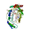
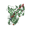
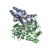
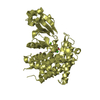
| Entry | Database: PDB / ID: 6heh | |||||||||
|---|---|---|---|---|---|---|---|---|---|---|
| Title | Structure of the catalytic domain of USP28 (insertion deleted) | |||||||||
 Components Components | Ubiquitin carboxyl-terminal hydrolase 28,Ubiquitin carboxyl-terminal hydrolase 28 | |||||||||
 Keywords Keywords |  HYDROLASE / Ubiquitin / USP / Ubiquitin-specific protease / DUB / Deubiquitinase / protease / isopeptidase / USP28 HYDROLASE / Ubiquitin / USP / Ubiquitin-specific protease / DUB / Deubiquitinase / protease / isopeptidase / USP28 | |||||||||
| Function / homology |  Function and homology information Function and homology informationprotein deubiquitination involved in ubiquitin-dependent protein catabolic process / deubiquitinase activity / response to ionizing radiation / protein deubiquitination / intrinsic apoptotic signaling pathway in response to DNA damage by p53 class mediator / DNA damage checkpoint signaling / regulation of protein stability / cellular response to UV / ubiquitinyl hydrolase 1 / cell population proliferation ...protein deubiquitination involved in ubiquitin-dependent protein catabolic process / deubiquitinase activity / response to ionizing radiation / protein deubiquitination / intrinsic apoptotic signaling pathway in response to DNA damage by p53 class mediator / DNA damage checkpoint signaling / regulation of protein stability / cellular response to UV / ubiquitinyl hydrolase 1 / cell population proliferation / cysteine-type deubiquitinase activity / nuclear body / Ub-specific processing proteases / DNA repair / DNA damage response / protein-containing complex / nucleoplasm / nucleus / cytosolSimilarity search - Function | |||||||||
| Biological species |  Homo sapiens (human) Homo sapiens (human) | |||||||||
| Method | X-RAY DIFFRACTION / SYNCHROTRON / MOLECULAR REPLACEMENT / Resolution: 2.26 Å | |||||||||
 Authors Authors | Gersch, M. / Komander, D. | |||||||||
| Funding support |  United Kingdom, 2items United Kingdom, 2items
| |||||||||
 Citation Citation | Journal: Mol.Cell / Year: 2019 Title: Distinct USP25 and USP28 Oligomerization States Regulate Deubiquitinating Activity. Authors: Gersch, M. / Wagstaff, J.L. / Toms, A.V. / Graves, B. / Freund, S.M.V. / Komander, D. | |||||||||
| History |
|
- Structure visualization
| Structure viewer | Molecule: MolmilJmol/JSmol |
|---|
- Downloads & links
-Download
| PDBx/mmCIF format | 6heh.cif.gz | 88.1 KB | Display | PDBx/mmCIF format |
|---|---|---|---|---|
| PDB format | pdb6heh.ent.gz | 64.7 KB | Display | PDB format |
| PDBx/mmJSON format | 6heh.json.gz | Tree view | PDBx/mmJSON format | |
| Others |  Other downloads Other downloads |
-Validation report
| Arichive directory | https://data.pdbj.org/pub/pdb/validation_reports/he/6hehftp://data.pdbj.org/pub/pdb/validation_reports/he/6heh | HTTPS FTP |
|---|
-Related structure data
| Related structure data | 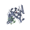 6heiSC 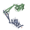 6hejC 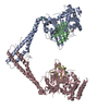 6hekC  6helC 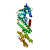 6hemC S: Starting model for refinement C: citing same article ( |
|---|---|
| Similar structure data |


-Links
 PDBj
PDBj

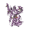
- Assembly
| Deposited unit | 
| ||||||||
|---|---|---|---|---|---|---|---|---|---|
| 1 |
| ||||||||
| Unit cell |
| ||||||||
| Components on special symmetry positions |
|
-Components
| #1: Protein | Mass: 44586.691 Da / Num. of mol.: 1 Mutation: residues 400-579 replaced by GSGSGS,residues 400-579 replaced by GSGSGS,residues 400-579 replaced by GSGSGS,residues 400-579 replaced by GSGSGS Source method: isolated from a genetically manipulated source Source: (gene. exp.) Homo sapiens (human) / Gene: USP28, KIAA1515 / Production host:  Escherichia coli BL21(DE3) (bacteria) / Variant (production host): Rosetta2 pLacI / References: UniProt: Q96RU2, ubiquitinyl hydrolase 1 Escherichia coli BL21(DE3) (bacteria) / Variant (production host): Rosetta2 pLacI / References: UniProt: Q96RU2, ubiquitinyl hydrolase 1 |
|---|---|
| #2: Chemical | ChemComp-EDO / Ethylene glycol  Mass: 62.068 Da / Num. of mol.: 1 / Source method: obtained synthetically / Formula: C2H6O2 Mass: 62.068 Da / Num. of mol.: 1 / Source method: obtained synthetically / Formula: C2H6O2 |
| #3: Chemical | ChemComp-CL / Chloride  Mass: 35.453 Da / Num. of mol.: 1 / Source method: obtained synthetically / Formula: Cl Mass: 35.453 Da / Num. of mol.: 1 / Source method: obtained synthetically / Formula: Cl |
| #4: Water | ChemComp-HOH / Water Mass: 18.015 Da / Num. of mol.: 128 / Source method: isolated from a natural source / Formula: H2O Mass: 18.015 Da / Num. of mol.: 128 / Source method: isolated from a natural source / Formula: H2O |
-Experimental details
-Experiment
| Experiment | Method: X-RAY DIFFRACTION / Number of used crystals: 1 |
|---|
- Sample preparation
| Crystal | Density Matthews: 3.16 Å3/Da / Density % sol: 61.1 % |
|---|---|
| Crystal grow | Temperature: 291 K / Method: vapor diffusion, sitting drop / pH: 6.6 Details: 12% (w/v) PEG 8000, 100 mM sodium chloride, 200 mM lithium sulfate, 100 mM MES pH 6.6 |
-Data collection
| Diffraction | Mean temperature: 100 K |
|---|---|
| Diffraction source | Source: SYNCHROTRON / Site: ESRF  / Beamline: ID30B / Wavelength: 0.9686 Å / Beamline: ID30B / Wavelength: 0.9686 Å |
| Detector | Type: DECTRIS PILATUS3 6M / Detector: PIXEL / Date: Jul 8, 2016 |
| Radiation | Protocol: SINGLE WAVELENGTH / Monochromatic (M) / Laue (L): M / Scattering type: x-ray |
| Radiation wavelength | Wavelength: 0.9686 Å / Relative weight: 1 |
| Reflection | Resolution: 2.26→50.55 Å / Num. obs: 27219 / % possible obs: 99.8 % / Redundancy: 8.6 % / Biso Wilson estimate: 44 Å2 / CC1/2: 0.999 / Rmerge(I) obs: 0.065 / Rrim(I) all: 0.068 / Net I/σ(I): 19.8 |
| Reflection shell | Resolution: 2.26→2.34 Å / Redundancy: 7.5 % / Rmerge(I) obs: 0.699 / Mean I/σ(I) obs: 2.7 / Num. unique obs: 2698 / CC1/2: 0.827 / Rrim(I) all: 0.752 / % possible all: 99.2 |
- Processing
| Software |
| |||||||||||||||||||||||||||||||||||||||||||||||||||||||||||||||||||||||||||||
|---|---|---|---|---|---|---|---|---|---|---|---|---|---|---|---|---|---|---|---|---|---|---|---|---|---|---|---|---|---|---|---|---|---|---|---|---|---|---|---|---|---|---|---|---|---|---|---|---|---|---|---|---|---|---|---|---|---|---|---|---|---|---|---|---|---|---|---|---|---|---|---|---|---|---|---|---|---|---|
| Refinement | Method to determine structure: MOLECULAR REPLACEMENT Starting model: 6HEI Resolution: 2.26→50.55 Å / SU ML: 0.21 / Cross valid method: FREE R-VALUE / σ(F): 1.35 / Phase error: 21.71
| |||||||||||||||||||||||||||||||||||||||||||||||||||||||||||||||||||||||||||||
| Solvent computation | Shrinkage radii: 0.9 Å / VDW probe radii: 1.11 Å | |||||||||||||||||||||||||||||||||||||||||||||||||||||||||||||||||||||||||||||
| Refinement step | Cycle: LAST / Resolution: 2.26→50.55 Å
| |||||||||||||||||||||||||||||||||||||||||||||||||||||||||||||||||||||||||||||
| Refine LS restraints |
| |||||||||||||||||||||||||||||||||||||||||||||||||||||||||||||||||||||||||||||
| LS refinement shell |
|
