Movie
Movie Controller

[English] 日本語
 Yorodumi
- PDB-7tqs: Coxsackievirus A21 capsid subdomain in complex with mouse polyclo... -
+ Open data

- Basic information
| Entry | Database: PDB / ID: 7tqs | ||||||
|---|---|---|---|---|---|---|---|
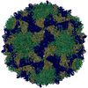
| Title | Coxsackievirus A21 capsid subdomain in complex with mouse polyclonal antibody pAbC-3 | ||||||
 Components Components |
| ||||||
 Keywords Keywords | VIRUS/IMMUNE SYSTEM / polyclonal antibodies / immune complex / cryoEM / VIRUS-IMMUNE SYSTEM complex | ||||||
| Function / homology |  Function and homology information Function and homology informationpicornain 2A / symbiont-mediated suppression of host mRNA export from nucleus / ribonucleoside triphosphate phosphatase activity / symbiont genome entry into host cell via pore formation in plasma membrane / picornain 3C / T=pseudo3 icosahedral viral capsid / host cell cytoplasmic vesicle membrane / cytoplasmic vesicle membrane / endocytosis involved in viral entry into host cell / nucleoside-triphosphate phosphatase ...picornain 2A / symbiont-mediated suppression of host mRNA export from nucleus / ribonucleoside triphosphate phosphatase activity / symbiont genome entry into host cell via pore formation in plasma membrane / picornain 3C / T=pseudo3 icosahedral viral capsid / host cell cytoplasmic vesicle membrane / cytoplasmic vesicle membrane / endocytosis involved in viral entry into host cell / nucleoside-triphosphate phosphatase / protein complex oligomerization / monoatomic ion channel activity / RNA helicase activity / symbiont-mediated suppression of host innate immune response / induction by virus of host autophagy / cysteine-type endopeptidase activity / RNA-directed RNA polymerase / viral RNA genome replication / virus-mediated perturbation of host defense response / RNA-dependent RNA polymerase activity / DNA-templated transcription / host cell nucleus / virion attachment to host cell / structural molecule activity / proteolysis / RNA binding / ATP binding / metal ion binding Similarity search - Function | ||||||
| Biological species |   Coxsackievirus A21 Coxsackievirus A21 | ||||||
| Method | ELECTRON MICROSCOPY / single particle reconstruction / cryo EM / Resolution: 3.9 Å | ||||||
 Authors Authors | Antanasijevic, A. / Ward, A.B. | ||||||
| Funding support | 1items
| ||||||
 Citation Citation | Journal: PNAS Nexus / Year: 2022 Title: High-resolution structural analysis of enterovirus-reactive polyclonal antibodies in complex with whole virions. Authors: Aleksandar Antanasijevic / Autumn J Schulze / Vijay S Reddy / Andrew B Ward /  Abstract: Non-polio enteroviruses (NPEVs) cause serious illnesses in young children and neonates, including aseptic meningitis, encephalitis, and inflammatory muscle disease, among others. While over 100 ...Non-polio enteroviruses (NPEVs) cause serious illnesses in young children and neonates, including aseptic meningitis, encephalitis, and inflammatory muscle disease, among others. While over 100 serotypes have been described to date, vaccine only exists for EV-A71. Efforts toward rationally designed pan-NPEV vaccines would greatly benefit from structural biology methods for rapid and comprehensive evaluation of vaccine candidates and elicited antibody responses. Toward this goal, we introduced a cryo-electron-microscopy-based approach for structural analysis of virus- or vaccine-elicited polyclonal antibodies (pAbs) in complex with whole NPEV virions. We demonstrated the feasibility using coxsackievirus A21 and reconstructed five structurally distinct pAbs bound to the virus. The pAbs targeted two immunodominant epitopes, one overlapping with the receptor binding site. These results demonstrate that our method can be applied to map broad-spectrum polyclonal immune responses against intact virions and define potentially cross-reactive epitopes. | ||||||
| History |
|
- Structure visualization
| Structure viewer | Molecule: MolmilJmol/JSmol |
|---|
- Downloads & links
-Download
| PDBx/mmCIF format | 7tqs.cif.gz | 824.2 KB | Display | PDBx/mmCIF format |
|---|---|---|---|---|
| PDB format | pdb7tqs.ent.gz | 674.6 KB | Display | PDB format |
| PDBx/mmJSON format | 7tqs.json.gz | Tree view | PDBx/mmJSON format | |
| Others |  Other downloads Other downloads |
-Validation report
| Summary document | 7tqs_validation.pdf.gz | 1.8 MB | Display | wwPDB validaton report |
|---|---|---|---|---|
| Full document | 7tqs_full_validation.pdf.gz | 1.9 MB | Display | |
| Data in XML | 7tqs_validation.xml.gz | 115.1 KB | Display | |
| Data in CIF | 7tqs_validation.cif.gz | 182.3 KB | Display | |
| Arichive directory | https://data.pdbj.org/pub/pdb/validation_reports/tq/7tqsftp://data.pdbj.org/pub/pdb/validation_reports/tq/7tqs | HTTPS FTP |
-Related structure data
| Related structure data | 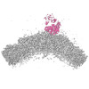 26068MC 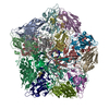 7tqtC 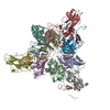 7tquC C: citing same article ( M: map data used to model this data |
|---|---|
| Similar structure data |
-Links
 PDBj
PDBj


- Assembly
| Deposited unit | 
|
|---|---|
| 1 |
|
-Components
-Protein , 6 types, 22 molecules LHaeimqbfjnrcgkosdhlpt
| #1: Protein | Mass: 8188.084 Da / Num. of mol.: 1 / Source method: isolated from a natural source / Details: Light Chain of the polyclonal antibody pAbC-3 / Source: (natural) | ||||||
|---|---|---|---|---|---|---|---|
| #2: Protein | Mass: 9039.134 Da / Num. of mol.: 1 / Source method: isolated from a natural source / Details: Light Chain of the polyclonal antibody pAbC-3 / Source: (natural) | ||||||
| #3: Protein | Mass: 33234.238 Da / Num. of mol.: 5 / Source method: isolated from a natural source / Details: VP1 protein of the coxsackievirus A21 capsid / Source: (natural) Coxsackievirus A21References: UniProt: Q7T7N6, picornain 2A, nucleoside-triphosphate phosphatase, picornain 3C, RNA-directed RNA polymerase #4: Protein | Mass: 29918.537 Da / Num. of mol.: 5 / Source method: isolated from a natural source / Details: VP2 protein of the coxsackievirus A21 capsid / Source: (natural) Coxsackievirus A21References: UniProt: Q7T7N6, picornain 2A, nucleoside-triphosphate phosphatase, picornain 3C, RNA-directed RNA polymerase #5: Protein | Mass: 26596.703 Da / Num. of mol.: 5 / Source method: isolated from a natural source / Details: VP3 protein of the coxsackievirus A21 capsid / Source: (natural) Coxsackievirus A21References: UniProt: Q71LY2, picornain 2A, nucleoside-triphosphate phosphatase, picornain 3C, RNA-directed RNA polymerase #6: Protein | Mass: 7442.118 Da / Num. of mol.: 5 / Source method: isolated from a natural source / Details: VP4 protein of the coxsackievirus A21 capsid / Source: (natural) Coxsackievirus A21References: UniProt: Q7T7N6, picornain 2A, nucleoside-triphosphate phosphatase, picornain 3C, RNA-directed RNA polymerase |
-Non-polymers , 1 types, 5 molecules 
| #7: Chemical | ChemComp-MYR /  Mass: 228.371 Da / Num. of mol.: 5 / Source method: obtained synthetically / Formula: C14H28O2 Mass: 228.371 Da / Num. of mol.: 5 / Source method: obtained synthetically / Formula: C14H28O2 |
|---|
-Details
| Has ligand of interest | N |
|---|
-Experimental details
-Experiment
| Experiment | Method: ELECTRON MICROSCOPY |
|---|---|
| EM experiment | Aggregation state: PARTICLE / 3D reconstruction method: single particle reconstruction |
- Sample preparation
| Component | Name: Immune complex featuring subdomains of coxsackievirus A21 (CV-A21) viral particles in complex with mouse polyclonal antibodies Type: COMPLEX Details: The complexes were assembled by combining purified CV-A21 particles and polyclonal antibodies isolated from mice (as Fab fragments) Entity ID: #1-#6 / Source: MULTIPLE SOURCES | |||||||||||||||
|---|---|---|---|---|---|---|---|---|---|---|---|---|---|---|---|---|
| Molecular weight | Experimental value: NO | |||||||||||||||
| Source (natural) |
| |||||||||||||||
| Buffer solution | pH: 7.4 / Details: TBS, 0.2um filtered | |||||||||||||||
| Buffer component |
| |||||||||||||||
| Specimen | Conc.: 2.5 mg/ml / Embedding applied: NO / Shadowing applied: NO / Staining applied: NO / Vitrification applied: YES Details: The complex was generated by incubation of mouse polyclonal antibodies with recombinantly expressed CV-A21 virions and subsequent SEC purification. | |||||||||||||||
| Specimen support | Grid material: COPPER / Grid mesh size: 400 divisions/in. / Grid type: Quantifoil R2/1 | |||||||||||||||
| Vitrification | Instrument: FEI VITROBOT MARK IV / Cryogen name: ETHANE / Humidity: 10 % / Chamber temperature: 283 K / Details: 3-7s blot time |
- Electron microscopy imaging
| Experimental equipment |  Model: Titan Krios / Image courtesy: FEI Company |
|---|---|
| Microscopy | Model: FEI TITAN KRIOS |
| Electron gun | Electron source:  FIELD EMISSION GUN / Accelerating voltage: 300 kV / Illumination mode: FLOOD BEAM FIELD EMISSION GUN / Accelerating voltage: 300 kV / Illumination mode: FLOOD BEAM |
| Electron lens | Mode: BRIGHT FIELD / Nominal magnification: 130000 X / Nominal defocus max: 1800 nm / Nominal defocus min: 800 nm / Cs: 2.7 mm / C2 aperture diameter: 70 µm / Alignment procedure: COMA FREE |
| Specimen holder | Cryogen: NITROGEN / Specimen holder model: FEI TITAN KRIOS AUTOGRID HOLDER |
| Image recording | Average exposure time: 9 sec. / Electron dose: 50 e/Å2 / Detector mode: COUNTING / Film or detector model: GATAN K2 SUMMIT (4k x 4k) / Num. of grids imaged: 2 / Num. of real images: 3862 |
| Image scans | Sampling size: 5 µm / Movie frames/image: 45 |
- Processing
| EM software |
| ||||||||||||||||||||||||||||||||||||||||
|---|---|---|---|---|---|---|---|---|---|---|---|---|---|---|---|---|---|---|---|---|---|---|---|---|---|---|---|---|---|---|---|---|---|---|---|---|---|---|---|---|---|
| CTF correction | Type: PHASE FLIPPING ONLY | ||||||||||||||||||||||||||||||||||||||||
| Particle selection | Num. of particles selected: 22937 Details: Cryosparc template picker. Following the 2D classification and symmetry expansion the number of particles was 1334700. Symmetry-expanded particles were used for 3D classification and refinement. | ||||||||||||||||||||||||||||||||||||||||
| Symmetry | Point symmetry: C1 (asymmetric) | ||||||||||||||||||||||||||||||||||||||||
| 3D reconstruction | Resolution: 3.9 Å / Resolution method: FSC 0.143 CUT-OFF / Num. of particles: 29282 / Algorithm: BACK PROJECTION / Details: 19748 symmetry expanded particles. / Num. of class averages: 1 / Symmetry type: POINT | ||||||||||||||||||||||||||||||||||||||||
| Atomic model building | Protocol: RIGID BODY FIT / Space: REAL | ||||||||||||||||||||||||||||||||||||||||
| Atomic model building | PDB-ID: 1Z7S Accession code: 1Z7S / Source name: PDB / Type: experimental model |