 Movie
Movie Controller

[English] 日本語
 Yorodumi
- EMDB-26065: nsEM maps of coxsackievirus A21 viral particle alone and in compl... -
+ Open data

- Basic information
| Entry |  | |||||||||
|---|---|---|---|---|---|---|---|---|---|---|
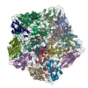
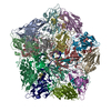
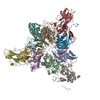
| Title | nsEM maps of coxsackievirus A21 viral particle alone and in complex with mouse polyclonal antibodies | |||||||||
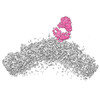
 Map data Map data | nsEM map of CV-A21 viral particle in complex with mouse polyclonal antibodies (antigen-specific) | |||||||||
 Sample Sample |
| |||||||||
 Keywords Keywords |  polyclonal antibodies / immune complex / negative stain EM / VIRUS / VIRUS-IMMUNE SYSTEM complex polyclonal antibodies / immune complex / negative stain EM / VIRUS / VIRUS-IMMUNE SYSTEM complex | |||||||||
| Biological species |  Coxsackievirus A21 Coxsackievirus A21 | |||||||||
| Method | single particle reconstruction / negative staining / Resolution: 15.0 Å | |||||||||
 Authors Authors | Antanasijevic A / Ward AB | |||||||||
| Funding support | 1 items
| |||||||||
 Citation Citation | Journal: PNAS Nexus / Year: 2022 Title: High-resolution structural analysis of enterovirus-reactive polyclonal antibodies in complex with whole virions. Authors: Aleksandar Antanasijevic / Autumn J Schulze / Vijay S Reddy / Andrew B Ward /  Abstract: Non-polio enteroviruses (NPEVs) cause serious illnesses in young children and neonates, including aseptic meningitis, encephalitis, and inflammatory muscle disease, among others. While over 100 ...Non-polio enteroviruses (NPEVs) cause serious illnesses in young children and neonates, including aseptic meningitis, encephalitis, and inflammatory muscle disease, among others. While over 100 serotypes have been described to date, vaccine only exists for EV-A71. Efforts toward rationally designed pan-NPEV vaccines would greatly benefit from structural biology methods for rapid and comprehensive evaluation of vaccine candidates and elicited antibody responses. Toward this goal, we introduced a cryo-electron-microscopy-based approach for structural analysis of virus- or vaccine-elicited polyclonal antibodies (pAbs) in complex with whole NPEV virions. We demonstrated the feasibility using coxsackievirus A21 and reconstructed five structurally distinct pAbs bound to the virus. The pAbs targeted two immunodominant epitopes, one overlapping with the receptor binding site. These results demonstrate that our method can be applied to map broad-spectrum polyclonal immune responses against intact virions and define potentially cross-reactive epitopes. | |||||||||
| History |
|
- Structure visualization
| Supplemental images |
|---|

- Downloads & links
-EMDB archive
| Map data | emd_26065.map.gz | 97.1 MB |  EMDB map data format EMDB map data format | |
|---|---|---|---|---|
| Header (meta data) | emd-26065-v30.xmlemd-26065.xml | 16.8 KB 16.8 KB | Display Display | EMDB header |
| Images | 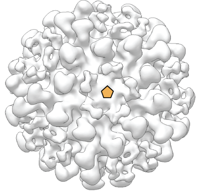 emd_26065.png emd_26065.png | 166.3 KB | ||
| Filedesc metadata | emd-26065.cif.gz | 4.3 KB | ||
| Others | emd_26065_additional_1.map.gzemd_26065_additional_2.map.gz | 33.6 MB 34.4 MB | ||
| Archive directory |  http://ftp.pdbj.org/pub/emdb/structures/EMD-26065ftp://ftp.pdbj.org/pub/emdb/structures/EMD-26065 http://ftp.pdbj.org/pub/emdb/structures/EMD-26065ftp://ftp.pdbj.org/pub/emdb/structures/EMD-26065 | HTTPS FTP |
-Related structure data
-Links
| EMDB pages | EMDB (EBI/PDBe) / EMDataResource |
|---|
-Map
| File | Download / File: emd_26065.map.gz / Format: CCP4 / Size: 125 MB / Type: IMAGE STORED AS FLOATING POINT NUMBER (4 BYTES) | ||||||||||||||||||||
|---|---|---|---|---|---|---|---|---|---|---|---|---|---|---|---|---|---|---|---|---|---|
| Annotation | nsEM map of CV-A21 viral particle in complex with mouse polyclonal antibodies (antigen-specific) | ||||||||||||||||||||
| Voxel size | X=Y=Z: 1.77 Å | ||||||||||||||||||||
| Density |
| ||||||||||||||||||||
| Symmetry | Space group: 1 | ||||||||||||||||||||
| Details | EMDB XML:
|
-Supplemental data
-Additional map: nsEM map of CV-A21 viral particle
| File | emd_26065_additional_1.map | ||||||||||||
|---|---|---|---|---|---|---|---|---|---|---|---|---|---|
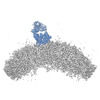
| Annotation | nsEM map of CV-A21 viral particle | ||||||||||||
| Projections & Slices |
| ||||||||||||
| Density Histograms |
 Z
Z Y
Y X
X


-Additional map: nsEM map of CV-A21 viral particle in complex...
| File | emd_26065_additional_2.map | ||||||||||||
|---|---|---|---|---|---|---|---|---|---|---|---|---|---|
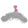
| Annotation | nsEM map of CV-A21 viral particle in complex with mouse polyclonal antibodies (non-specific) | ||||||||||||
| Projections & Slices |
| ||||||||||||
| Density Histograms |


- Sample components
-Entire : Immune complexes featuring coxsackievirus A21 (CV-A21) viral part...
| Entire | Name: Immune complexes featuring coxsackievirus A21 (CV-A21) viral particles by themselves or in complex with mouse polyclonal antibodies (reconstructed by nsEM) |
|---|---|
| Components |
|
-Supramolecule #1: Immune complexes featuring coxsackievirus A21 (CV-A21) viral part...
| Supramolecule | Name: Immune complexes featuring coxsackievirus A21 (CV-A21) viral particles by themselves or in complex with mouse polyclonal antibodies (reconstructed by nsEM) type: complex / ID: 1 / Parent: 0 Details: The 3 submitted maps feature nsEM reconstructions of: 1) CV-A21 in complex with mouse polyclonal antibodies (antigen-specific) 2) CV-A21 in complex with mouse polyclonal antibodies (non-specific) 3) CV-A21 alone |
|---|---|
| Source (natural) | Organism: Coxsackievirus A21 |
-Experimental details
-Structure determination
| Method | negative staining |
|---|---|
 Processing Processing | single particle reconstruction |
| Aggregation state | particle |
-Sample preparation
| Concentration | 0.1 mg/mL | |||||||||
|---|---|---|---|---|---|---|---|---|---|---|
| Buffer | pH: 7.4 Component:
Details: TBS, 0.2um filtered | |||||||||
| Staining | Type: NEGATIVE / Material: Uranyl formate / Details: The samples were stained with UF for 60s. | |||||||||
| Grid | Model: Homemade / Material: COPPER / Mesh: 400 / Support film - Material: CARBON / Pretreatment - Type: PLASMA CLEANING / Pretreatment - Time: 30 sec. | |||||||||
| Details | The complex was generated by incubation of mouse polyclonal antibodies with recombinantly expressed CV-A21 virions and subsequent SEC purification. |
- Electron microscopy
| Microscope | FEI TECNAI F20 |
|---|---|
| Electron beam | Acceleration voltage: 200 kV / Electron source: FIELD EMISSION GUN |
| Electron optics | C2 aperture diameter: 70.0 µm / Illumination mode: FLOOD BEAM / Imaging mode: BRIGHT FIELDBright-field microscopy / Cs: 2.2 mm / Nominal defocus max: 1.5 µm / Nominal defocus min: 1.5 µm / Nominal magnification: 62000 |
| Sample stage | Specimen holder model: SIDE ENTRY, EUCENTRIC |
| Image recording | Film or detector model: TVIPS TEMCAM-F416 (4k x 4k) / Average electron dose: 25.0 e/Å2 |
| Experimental equipment |  Model: Tecnai F20 / Image courtesy: FEI Company |
-Image processing
| Particle selection | Number selected: 6224 / Details: DoG Picker in Appion |
|---|---|
| Startup model | Type of model: OTHER / Details: Low-pass filtered map of coxsackievirus particle |
| Initial angle assignment | Type: OTHER / Software - Name: RELION (ver. 3.0) / Details: Relion, regularized likelihood |
| Final 3D classification | Number classes: 4 / Software - Name: RELION (ver. 3.0) |
| Final angle assignment | Type: OTHER / Software - Name: RELION (ver. 3.0) / Details: Relion, regularized likelihood |
| Final reconstruction | Applied symmetry - Point group: I (icosahedral) / Algorithm: BACK PROJECTION / Resolution.type: BY AUTHOR / Resolution: 15.0 Å / Resolution method: FSC 0.143 CUT-OFF / Software - Name: RELION (ver. 3.0) / Number images used: 6224 |
