 Movie
Movie Controller

[English] 日本語
 Yorodumi
- PDB-7tda: Crystal structure of the E. coli thiM riboswitch in complex with ... -
+ Open data

- Basic information
| Entry | Database: PDB / ID: 7tda | ||||||
|---|---|---|---|---|---|---|---|
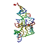
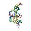
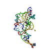
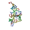
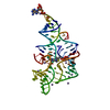
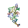
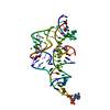
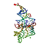
| Title | Crystal structure of the E. coli thiM riboswitch in complex with thiamine pyrophosphate, manganese ions | ||||||
 Components Components | thiM TPP riboswitch RNA (80-MER) | ||||||
 Keywords Keywords |  RNA RNA | ||||||
| Function / homology | : / THIAMINE DIPHOSPHATE / RNA / RNA (> 10) Function and homology information Function and homology information | ||||||
| Biological species |  Escherichia coli (E. coli) Escherichia coli (E. coli) | ||||||
| Method | X-RAY DIFFRACTION / SYNCHROTRON / MOLECULAR REPLACEMENT / Resolution: 2.25 Å | ||||||
 Authors Authors | Nuthanakanti, A. / Serganov, A. | ||||||
| Funding support |  United States, 1items United States, 1items
| ||||||
 Citation Citation | Journal: Acs Chem.Biol. / Year: 2022 Title: Subsite Ligand Recognition and Cooperativity in the TPP Riboswitch: Implications for Fragment-Linking in RNA Ligand Discovery. Authors: Zeller, M.J. / Nuthanakanti, A. / Li, K. / Aube, J. / Serganov, A. / Weeks, K.M. | ||||||
| History |
|
- Structure visualization
| Structure viewer | Molecule: MolmilJmol/JSmol |
|---|
- Downloads & links
-Download
| PDBx/mmCIF format | 7tda.cif.gz | 60.2 KB | Display | PDBx/mmCIF format |
|---|---|---|---|---|
| PDB format | pdb7tda.ent.gz | 40.6 KB | Display | PDB format |
| PDBx/mmJSON format | 7tda.json.gz | Tree view | PDBx/mmJSON format | |
| Others |  Other downloads Other downloads |
-Validation report
| Arichive directory | https://data.pdbj.org/pub/pdb/validation_reports/td/7tdaftp://data.pdbj.org/pub/pdb/validation_reports/td/7tda | HTTPS FTP |
|---|
-Related structure data
| Related structure data | 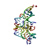 7td7C  7tdbC 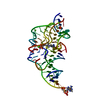 7tdcC  2hojS S: Starting model for refinement C: citing same article ( |
|---|---|
| Similar structure data |


-Links
 PDBj
PDBj


- Assembly
| Deposited unit | 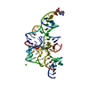
| ||||||||
|---|---|---|---|---|---|---|---|---|---|
| 1 |
| ||||||||
| Unit cell |
|
-Components
| #1: RNA chain | Mass: 26860.900 Da / Num. of mol.: 1 / Source method: obtained synthetically / Source: (synth.) Escherichia coli (E. coli) | ||||||
|---|---|---|---|---|---|---|---|
| #2: Chemical | ChemComp-TPP / Thiamine pyrophosphate  Mass: 425.314 Da / Num. of mol.: 1 / Source method: obtained synthetically / Formula: C12H19N4O7P2S / Feature type: SUBJECT OF INVESTIGATION Mass: 425.314 Da / Num. of mol.: 1 / Source method: obtained synthetically / Formula: C12H19N4O7P2S / Feature type: SUBJECT OF INVESTIGATION | ||||||
| #3: Chemical | ChemComp-MG /   Mass: 24.305 Da / Num. of mol.: 6 / Source method: obtained synthetically / Formula: Mg Mass: 24.305 Da / Num. of mol.: 6 / Source method: obtained synthetically / Formula: Mg#4: Chemical | ChemComp-MN /   Mass: 54.938 Da / Num. of mol.: 9 / Source method: obtained synthetically / Formula: Mn Mass: 54.938 Da / Num. of mol.: 9 / Source method: obtained synthetically / Formula: Mn#5: Water | ChemComp-HOH / | Water Mass: 18.015 Da / Num. of mol.: 33 / Source method: isolated from a natural source / Formula: H2O Mass: 18.015 Da / Num. of mol.: 33 / Source method: isolated from a natural source / Formula: H2OHas ligand of interest | Y | |
-Experimental details
-Experiment
| Experiment | Method: X-RAY DIFFRACTION / Number of used crystals: 1 |
|---|
- Sample preparation
| Crystal | Density Matthews: 2.33 Å3/Da / Density % sol: 47.29 % / Description: Rod-shaped crystals |
|---|---|
| Crystal grow | Temperature: 291 K / Method: vapor diffusion, hanging drop Details: RNA (0.15 mM) was incubated in a buffer containing 5 mM Tris-HCl, pH 8.0, 3 mM MgCl2, 10 mM NaCl, 0.1 M KCl, and 0.5 mM spermine with 0.5 mM of TPP. the reservoir solution was 50 mM Bis-Tris ...Details: RNA (0.15 mM) was incubated in a buffer containing 5 mM Tris-HCl, pH 8.0, 3 mM MgCl2, 10 mM NaCl, 0.1 M KCl, and 0.5 mM spermine with 0.5 mM of TPP. the reservoir solution was 50 mM Bis-Tris HCl, pH 6.5, 0.5 M NH4Cl, 10 mm MnCl2, and 30% (v/v) PEG2000 PH range: 6.0-6.5 |
-Data collection
| Diffraction | Mean temperature: 100 K / Serial crystal experiment: N |
|---|---|
| Diffraction source | Source: SYNCHROTRON / Site: NSLS-II / Beamline: 17-ID-2 / Wavelength: 0.9793 Å |
| Detector | Type: DECTRIS EIGER X 16M / Detector: PIXEL / Date: Feb 1, 2021 |
| Radiation | Protocol: SINGLE WAVELENGTH / Monochromatic (M) / Laue (L): M / Scattering type: x-ray |
| Radiation wavelength | Wavelength: 0.9793 Å / Relative weight: 1 |
| Reflection | Resolution: 2.25→30 Å / Num. obs: 12071 / % possible obs: 100 % / Redundancy: 12.4 % / CC1/2: 0.994 / Rmerge(I) obs: 0.064 / Rpim(I) all: 0.019 / Net I/σ(I): 82.7 |
| Reflection shell | Resolution: 2.25→2.29 Å / Redundancy: 9.6 % / Rmerge(I) obs: 1.297 / Mean I/σ(I) obs: 1 / Num. unique obs: 589 / CC1/2: 0.7 / Rpim(I) all: 0.437 / % possible all: 100 |
- Processing
| Software |
| ||||||||||||||||||||||||||||||||||||||||||||||||||||||||||||||||||||||||||||||||||||||||||||||||||||||||||||||||||||||||||||||
|---|---|---|---|---|---|---|---|---|---|---|---|---|---|---|---|---|---|---|---|---|---|---|---|---|---|---|---|---|---|---|---|---|---|---|---|---|---|---|---|---|---|---|---|---|---|---|---|---|---|---|---|---|---|---|---|---|---|---|---|---|---|---|---|---|---|---|---|---|---|---|---|---|---|---|---|---|---|---|---|---|---|---|---|---|---|---|---|---|---|---|---|---|---|---|---|---|---|---|---|---|---|---|---|---|---|---|---|---|---|---|---|---|---|---|---|---|---|---|---|---|---|---|---|---|---|---|---|
| Refinement | Method to determine structure: MOLECULAR REPLACEMENT Starting model: 2HOJ Resolution: 2.25→29.05 Å / SU ML: 0.42 / Cross valid method: FREE R-VALUE / σ(F): 1.34 / Phase error: 31.27 / Stereochemistry target values: ML
| ||||||||||||||||||||||||||||||||||||||||||||||||||||||||||||||||||||||||||||||||||||||||||||||||||||||||||||||||||||||||||||||
| Solvent computation | Shrinkage radii: 0.9 Å / VDW probe radii: 1.11 Å / Solvent model: FLAT BULK SOLVENT MODEL | ||||||||||||||||||||||||||||||||||||||||||||||||||||||||||||||||||||||||||||||||||||||||||||||||||||||||||||||||||||||||||||||
| Refinement step | Cycle: LAST / Resolution: 2.25→29.05 Å
| ||||||||||||||||||||||||||||||||||||||||||||||||||||||||||||||||||||||||||||||||||||||||||||||||||||||||||||||||||||||||||||||
| Refine LS restraints |
| ||||||||||||||||||||||||||||||||||||||||||||||||||||||||||||||||||||||||||||||||||||||||||||||||||||||||||||||||||||||||||||||
| LS refinement shell |
|
