 Movie
Movie Controller


+ Open data

- Basic information
| Entry | Database: PDB / ID: 7t8d | ||||||
|---|---|---|---|---|---|---|---|
| Title | Myocilin OLF mutant V449I | ||||||
 Components Components | Myocilin, C-terminal fragment | ||||||
 Keywords Keywords | CELL ADHESION / olfactomedin / beta-propeller | ||||||
| Function / homology |  Function and homology information Function and homology informationskeletal muscle hypertrophy / clustering of voltage-gated sodium channels / myosin light chain binding / non-canonical Wnt signaling pathway / myelination in peripheral nervous system / node of Ranvier / frizzled binding / negative regulation of stress fiber assembly / negative regulation of Rho protein signal transduction / positive regulation of mitochondrial depolarization ...skeletal muscle hypertrophy / clustering of voltage-gated sodium channels / myosin light chain binding / non-canonical Wnt signaling pathway / myelination in peripheral nervous system / node of Ranvier / frizzled binding / negative regulation of stress fiber assembly / negative regulation of Rho protein signal transduction / positive regulation of mitochondrial depolarization / negative regulation of cell-matrix adhesion / ERBB2-ERBB3 signaling pathway / positive regulation of focal adhesion assembly / regulation of MAPK cascade / fibronectin binding / rough endoplasmic reticulum / positive regulation of substrate adhesion-dependent cell spreading / positive regulation of stress fiber assembly / positive regulation of JNK cascade / bone development / mitochondrial intermembrane space / cilium / receptor tyrosine kinase binding / osteoblast differentiation / neuron projection development / cytoplasmic vesicle / collagen-containing extracellular matrix / mitochondrial outer membrane / mitochondrial inner membrane / positive regulation of phosphatidylinositol 3-kinase/protein kinase B signal transduction / positive regulation of cell migration / Golgi apparatus / endoplasmic reticulum / signal transduction / extracellular space / extracellular exosome / metal ion bindingSimilarity search - Function | ||||||
| Biological species |  Homo sapiens (human) Homo sapiens (human) | ||||||
| Method | X-RAY DIFFRACTION / SYNCHROTRON / MOLECULAR REPLACEMENT / Resolution: 1.38 Å | ||||||
 Authors Authors | Scelsi, H.S. / Barlow, B.M. / Lieberman, R.L. | ||||||
| Funding support |  United States, 1items United States, 1items
| ||||||
 Citation Citation | Journal: Dis Model Mech / Year: 2023 Title: Quantitative differentiation of benign and misfolded glaucoma-causing myocilin variants on the basis of protein thermal stability. Authors: Scelsi, H.F. / Hill, K.R. / Barlow, B.M. / Martin, M.D. / Lieberman, R.L. | ||||||
| History |
|
- Structure visualization
| Structure viewer | Molecule: MolmilJmol/JSmol |
|---|
- Downloads & links
-Download
| PDBx/mmCIF format | 7t8d.cif.gz | 89.7 KB | Display | PDBx/mmCIF format |
|---|---|---|---|---|
| PDB format | pdb7t8d.ent.gz | 52 KB | Display | PDB format |
| PDBx/mmJSON format | 7t8d.json.gz | Tree view | PDBx/mmJSON format | |
| Others |  Other downloads Other downloads |
-Validation report
| Arichive directory | https://data.pdbj.org/pub/pdb/validation_reports/t8/7t8dftp://data.pdbj.org/pub/pdb/validation_reports/t8/7t8d | HTTPS FTP |
|---|
-Related structure data
| Related structure data |  7sibC  7sijC  7sjtC  7sjuC  7sjvC  7sjwC  7skdC  7skeC 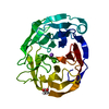 7skfC 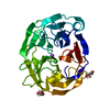 7skgC  6pkeS S: Starting model for refinement C: citing same article ( |
|---|---|
| Similar structure data |
-Links
 PDBj
PDBj- Assembly
| Deposited unit | 
| ||||||||||||
|---|---|---|---|---|---|---|---|---|---|---|---|---|---|
| 1 |
| ||||||||||||
| Unit cell |
|
-Components
| #1: Protein | / Myocilin 35 kDa N-terminal fragment Mass: 31211.008 Da / Num. of mol.: 1 / Mutation: V449I Source method: isolated from a genetically manipulated source Source: (gene. exp.) Homo sapiens (human) / Gene: MYOC, GLC1A, TIGR / Production host:  Escherichia coli (E. coli) / References: UniProt: Q99972 Escherichia coli (E. coli) / References: UniProt: Q99972 |
|---|---|
| #2: Chemical | ChemComp-CA /   Mass: 40.078 Da / Num. of mol.: 1 / Source method: obtained synthetically / Formula: Ca Mass: 40.078 Da / Num. of mol.: 1 / Source method: obtained synthetically / Formula: Ca |
| #3: Chemical | ChemComp-NA /   Mass: 22.990 Da / Num. of mol.: 1 / Source method: obtained synthetically / Formula: Na Mass: 22.990 Da / Num. of mol.: 1 / Source method: obtained synthetically / Formula: Na |
| #4: Chemical | ChemComp-GOL / Glycerol  Mass: 92.094 Da / Num. of mol.: 1 / Source method: obtained synthetically / Formula: C3H8O3 Mass: 92.094 Da / Num. of mol.: 1 / Source method: obtained synthetically / Formula: C3H8O3 |
| #5: Water | ChemComp-HOH / Water Mass: 18.015 Da / Num. of mol.: 216 / Source method: isolated from a natural source / Formula: H2O Mass: 18.015 Da / Num. of mol.: 216 / Source method: isolated from a natural source / Formula: H2O |
| Has ligand of interest | N |
-Experimental details
-Experiment
| Experiment | Method: X-RAY DIFFRACTION / Number of used crystals: 1 |
|---|
- Sample preparation
| Crystal | Density Matthews: 1.97 Å3/Da / Density % sol: 37.49 % |
|---|---|
| Crystal grow | Temperature: 289 K / Method: vapor diffusion, hanging drop / pH: 7.2 / Details: 0.05 M magnesium chloride, 10% PEG 8000 |
-Data collection
| Diffraction | Mean temperature: 273 K / Serial crystal experiment: N |
|---|---|
| Diffraction source | Source: SYNCHROTRON / Site: APS / Beamline: 22-ID / Wavelength: 1 Å |
| Detector | Type: DECTRIS EIGER X 16M / Detector: PIXEL / Date: Dec 9, 2021 |
| Radiation | Protocol: SINGLE WAVELENGTH / Monochromatic (M) / Laue (L): M / Scattering type: x-ray |
| Radiation wavelength | Wavelength: 1 Å / Relative weight: 1 |
| Reflection | Resolution: 1.38→35.69 Å / Num. obs: 50378 / % possible obs: 98.68 % / Redundancy: 6.7 % / Biso Wilson estimate: 9.74 Å2 / CC1/2: 0.998 / CC star: 0.999 / Rmerge(I) obs: 0.0661 / Rpim(I) all: 0.0274 / Rrim(I) all: 0.07168 / Net I/σ(I): 17.82 |
| Reflection shell | Resolution: 1.38→1.429 Å / Redundancy: 6.3 % / Rmerge(I) obs: 0.4149 / Mean I/σ(I) obs: 6.52 / Num. unique obs: 4924 / CC1/2: 0.935 / CC star: 0.983 / Rpim(I) all: 0.182 / Rrim(I) all: 0.4541 / % possible all: 97.45 |
- Processing
| Software |
| |||||||||||||||||||||||||||||||||||||||||||||||||||||||||||||||||||||||||||||||||||||||||||||||||||||||||
|---|---|---|---|---|---|---|---|---|---|---|---|---|---|---|---|---|---|---|---|---|---|---|---|---|---|---|---|---|---|---|---|---|---|---|---|---|---|---|---|---|---|---|---|---|---|---|---|---|---|---|---|---|---|---|---|---|---|---|---|---|---|---|---|---|---|---|---|---|---|---|---|---|---|---|---|---|---|---|---|---|---|---|---|---|---|---|---|---|---|---|---|---|---|---|---|---|---|---|---|---|---|---|---|---|---|---|
| Refinement | Method to determine structure: MOLECULAR REPLACEMENT Starting model: 6PKE Resolution: 1.38→35.69 Å / SU ML: 0.1173 / Cross valid method: FREE R-VALUE / σ(F): 1.39 / Phase error: 18.1856 Stereochemistry target values: GeoStd + Monomer Library + CDL v1.2
| |||||||||||||||||||||||||||||||||||||||||||||||||||||||||||||||||||||||||||||||||||||||||||||||||||||||||
| Solvent computation | Shrinkage radii: 0.9 Å / VDW probe radii: 1.11 Å / Solvent model: FLAT BULK SOLVENT MODEL | |||||||||||||||||||||||||||||||||||||||||||||||||||||||||||||||||||||||||||||||||||||||||||||||||||||||||
| Displacement parameters | Biso mean: 12.97 Å2 | |||||||||||||||||||||||||||||||||||||||||||||||||||||||||||||||||||||||||||||||||||||||||||||||||||||||||
| Refinement step | Cycle: LAST / Resolution: 1.38→35.69 Å
| |||||||||||||||||||||||||||||||||||||||||||||||||||||||||||||||||||||||||||||||||||||||||||||||||||||||||
| Refine LS restraints |
| |||||||||||||||||||||||||||||||||||||||||||||||||||||||||||||||||||||||||||||||||||||||||||||||||||||||||
| LS refinement shell |
|
