 Movie
Movie Controller

[English] 日本語
 Yorodumi
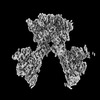
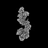
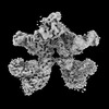
- PDB-7t4q: CryoEM structure of the HCMV Pentamer gH/gL/UL128/UL130/UL131A in... -
+ Open data

- Basic information
| Entry | Database: PDB / ID: 7t4q | ||||||
|---|---|---|---|---|---|---|---|
| Title | CryoEM structure of the HCMV Pentamer gH/gL/UL128/UL130/UL131A in complex with neutralizing fabs 2C12, 7I13 and 13H11 | ||||||
 Components Components |
| ||||||
 Keywords Keywords |  VIRAL PROTEIN/Immune System / Human Cytomegalovirus / glycoprotein complex / antibody complex / VIRAL PROTEIN / VIRAL PROTEIN-Immune System complex VIRAL PROTEIN/Immune System / Human Cytomegalovirus / glycoprotein complex / antibody complex / VIRAL PROTEIN / VIRAL PROTEIN-Immune System complex | ||||||
| Function / homology |  Function and homology information Function and homology informationhost cell endosome membrane / host cell Golgi apparatus / entry receptor-mediated virion attachment to host cell / symbiont entry into host cell / fusion of virus membrane with host plasma membrane / viral envelope / host cell plasma membrane / virion membrane / membraneSimilarity search - Function | ||||||
| Biological species |   Human betaherpesvirus 5 Human betaherpesvirus 5 Homo sapiens (human) Homo sapiens (human) | ||||||
| Method | ELECTRON MICROSCOPY / single particle reconstruction / cryo EM / Resolution: 2.9 Å | ||||||
 Authors Authors | Kschonsak, M. / Johnson, M.C. / Schelling, R. / Green, E.M. / Rouge, L. / Ho, H. / Patel, N. / Kilic, C. / Kraft, E. / Arthur, C.P. ...Kschonsak, M. / Johnson, M.C. / Schelling, R. / Green, E.M. / Rouge, L. / Ho, H. / Patel, N. / Kilic, C. / Kraft, E. / Arthur, C.P. / Rohou, A.L. / Comps-Agrar, L. / Martinez-Martin, N. / Perez, L. / Payandeh, J. / Ciferri, C. | ||||||
| Funding support | 1items
| ||||||
 Citation Citation | Journal: Sci Adv / Year: 2022 Title: Structural basis for HCMV Pentamer receptor recognition and antibody neutralization. Authors: Marc Kschonsak / Matthew C Johnson / Rachel Schelling / Evan M Green / Lionel Rougé / Hoangdung Ho / Nidhi Patel / Cem Kilic / Edward Kraft / Christopher P Arthur / Alexis L Rohou / ...Authors: Marc Kschonsak / Matthew C Johnson / Rachel Schelling / Evan M Green / Lionel Rougé / Hoangdung Ho / Nidhi Patel / Cem Kilic / Edward Kraft / Christopher P Arthur / Alexis L Rohou / Laetitia Comps-Agrar / Nadia Martinez-Martin / Laurent Perez / Jian Payandeh / Claudio Ciferri /   Abstract: Human cytomegalovirus (HCMV) represents the viral leading cause of congenital birth defects and uses the gH/gL/UL128-130-131A complex (Pentamer) to enter different cell types, including epithelial ...Human cytomegalovirus (HCMV) represents the viral leading cause of congenital birth defects and uses the gH/gL/UL128-130-131A complex (Pentamer) to enter different cell types, including epithelial and endothelial cells. Upon infection, Pentamer elicits the most potent neutralizing response against HCMV, representing a key vaccine candidate. Despite its relevance, the structural basis for Pentamer receptor recognition and antibody neutralization is largely unknown. Here, we determine the structures of Pentamer bound to neuropilin 2 (NRP2) and a set of potent neutralizing antibodies against HCMV. Moreover, we identify thrombomodulin (THBD) as a functional HCMV receptor and determine the structures of the Pentamer-THBD complex. Unexpectedly, both NRP2 and THBD also promote dimerization of Pentamer. Our results provide a framework for understanding HCMV receptor engagement, cell entry, antibody neutralization, and outline strategies for antiviral therapies against HCMV. | ||||||
| History |
|
- Structure visualization
| Structure viewer | Molecule: MolmilJmol/JSmol |
|---|
- Downloads & links
-Download
| PDBx/mmCIF format | 7t4q.cif.gz | 368.7 KB | Display | PDBx/mmCIF format |
|---|---|---|---|---|
| PDB format | pdb7t4q.ent.gz | 294.4 KB | Display | PDB format |
| PDBx/mmJSON format | 7t4q.json.gz | Tree view | PDBx/mmJSON format | |
| Others |  Other downloads Other downloads |
-Validation report
| Arichive directory | https://data.pdbj.org/pub/pdb/validation_reports/t4/7t4qftp://data.pdbj.org/pub/pdb/validation_reports/t4/7t4q | HTTPS FTP |
|---|
-Related structure data
| Related structure data | 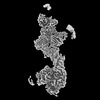 25685MC 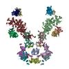 7t4rC 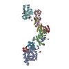 7t4sC M: map data used to model this data C: citing same article ( |
|---|---|
| Similar structure data |
-Links
 PDBj
PDBj

- Assembly
| Deposited unit | 
|
|---|---|
| 1 |
|
-Components
-Envelope glycoprotein ... , 3 types, 3 molecules ABD
| #1: Protein | Mass: 87311.273 Da / Num. of mol.: 1 Source method: isolated from a genetically manipulated source Source: (gene. exp.) Human betaherpesvirus 5 / Gene: UL75, gH / Production host: Homo sapiens (human) / References: UniProt: F5H9T3 |
|---|---|
| #2: Protein | Mass: 30846.492 Da / Num. of mol.: 1 Source method: isolated from a genetically manipulated source Source: (gene. exp.) Human betaherpesvirus 5 / Gene: UL115, gL / Production host: Homo sapiens (human) / References: UniProt: Q71DN9 |
| #4: Protein | Mass: 28866.811 Da / Num. of mol.: 1 Source method: isolated from a genetically manipulated source Source: (gene. exp.) Human betaherpesvirus 5 / Gene: UL130 / Production host: Homo sapiens (human) / References: UniProt: Q38M07 |
-Envelope protein ... , 2 types, 2 molecules CE
| #3: Protein | / UL128 Mass: 19746.023 Da / Num. of mol.: 1 Source method: isolated from a genetically manipulated source Source: (gene. exp.) Human betaherpesvirus 5 / Gene: UL128 / Production host: Homo sapiens (human) / References: UniProt: Q38LY2 |
|---|---|
| #5: Protein | / Protein UL131A / UL131A Mass: 15011.043 Da / Num. of mol.: 1 Source method: isolated from a genetically manipulated source Source: (gene. exp.) Human betaherpesvirus 5 / Gene: UL131A, HHV5wtgp112 / Production host: Homo sapiens (human) / References: UniProt: Q8AZ45 |
-Antibody , 6 types, 6 molecules FGHIJK
| #6: Antibody | Mass: 27001.770 Da / Num. of mol.: 1 Source method: isolated from a genetically manipulated source Source: (gene. exp.) Homo sapiens (human) / Production host:  Escherichia coli (E. coli) Escherichia coli (E. coli) |
|---|---|
| #7: Antibody | Mass: 25594.654 Da / Num. of mol.: 1 Source method: isolated from a genetically manipulated source Source: (gene. exp.) Homo sapiens (human) / Production host: Escherichia coli (E. coli) |
| #8: Antibody | Mass: 25844.900 Da / Num. of mol.: 1 Source method: isolated from a genetically manipulated source Source: (gene. exp.) Homo sapiens (human) / Production host: Escherichia coli (E. coli) |
| #9: Antibody | Mass: 27046.506 Da / Num. of mol.: 1 Source method: isolated from a genetically manipulated source Source: (gene. exp.) Homo sapiens (human) / Production host: Escherichia coli (E. coli) |
| #10: Antibody | Mass: 26600.086 Da / Num. of mol.: 1 Source method: isolated from a genetically manipulated source Source: (gene. exp.) Homo sapiens (human) / Production host: Escherichia coli (E. coli) |
| #11: Antibody | Mass: 25780.020 Da / Num. of mol.: 1 Source method: isolated from a genetically manipulated source Source: (gene. exp.) Homo sapiens (human) / Production host: Escherichia coli (E. coli) |
-Sugars , 1 types, 6 molecules 
| #12: Sugar | ChemComp-NAG / N-Acetylglucosamine Type: D-saccharide, beta linking / Mass: 221.208 Da / Num. of mol.: 6 / Source method: obtained synthetically / Formula: C8H15NO6 Type: D-saccharide, beta linking / Mass: 221.208 Da / Num. of mol.: 6 / Source method: obtained synthetically / Formula: C8H15NO6 |
|---|
-Details
| Has ligand of interest | N |
|---|
-Experimental details
-Experiment
| Experiment | Method: ELECTRON MICROSCOPY |
|---|---|
| EM experiment | Aggregation state: PARTICLE / 3D reconstruction method: single particle reconstruction |
- Sample preparation
| Component | Name: Pentameric complex of HCMV proteins gH, gL, UL128, UL130, UL131A bound to fabs of human neutralizing antibodies 2C12, 7I13 and 13H11 Type: COMPLEX / Entity ID: #1-#11 / Source: MULTIPLE SOURCES | |||||||||||||||
|---|---|---|---|---|---|---|---|---|---|---|---|---|---|---|---|---|
| Molecular weight | Value: 0.32 MDa / Experimental value: NO | |||||||||||||||
| Details of virus | Empty: NO / Enveloped: YES / Isolate: STRAIN / Type: VIRION | |||||||||||||||
| Buffer solution | pH: 7.5 | |||||||||||||||
| Buffer component |
| |||||||||||||||
| Specimen | Conc.: 1.77 mg/ml / Embedding applied: NO / Shadowing applied: NO / Staining applied: NO / Vitrification applied: YES / Details: The sample was monodisperse. | |||||||||||||||
| Specimen support | Details: The grid was incubated with a thiol reactive self-assembling reaction mixture of 4mM monothiolalkane(C11)PEG6-OH (11-mercaptoundecyl) hexaethyleneglycol, (SPT-0011P6, SensoPath Technologies, ...Details: The grid was incubated with a thiol reactive self-assembling reaction mixture of 4mM monothiolalkane(C11)PEG6-OH (11-mercaptoundecyl) hexaethyleneglycol, (SPT-0011P6, SensoPath Technologies, Inc., Bozeman, MT)[23]. Grids were incubated with this self-assembled, monolayer (SAM) solution for 24 hours. Prior to grid freezing, grids were removed from the SAM solution and rinsed with EtOH. Grid material: GOLD / Grid mesh size: 300 divisions/in. / Grid type: UltrAuFoil R0.6/1 | |||||||||||||||
| Vitrification | Instrument: LEICA EM GP / Cryogen name: ETHANE / Humidity: 100 % / Chamber temperature: 277.15 K / Details: blot for 3.5s before plunging |
- Electron microscopy imaging
| Experimental equipment |  Model: Titan Krios / Image courtesy: FEI Company |
|---|---|
| Microscopy | Model: FEI TITAN KRIOS |
| Electron gun | Electron source: FIELD EMISSION GUN / Accelerating voltage: 300 kV / Illumination mode: FLOOD BEAM |
| Electron lens | Mode: BRIGHT FIELDBright-field microscopy / Nominal magnification: 165000 X / Nominal defocus max: 1500 nm / Nominal defocus min: 500 nm / Cs: 2.7 mm |
| Specimen holder | Cryogen: NITROGEN / Specimen holder model: FEI TITAN KRIOS AUTOGRID HOLDER |
| Image recording | Average exposure time: 10 sec. / Electron dose: 58 e/Å2 / Detector mode: COUNTING / Film or detector model: GATAN K2 SUMMIT (4k x 4k) / Num. of grids imaged: 1 / Num. of real images: 18326 Details: Images were collected in movie-mode at 5 frames/second. |
| Image scans | Movie frames/image: 50 |
- Processing
| Software | Name: PHENIX / Version: 1.19.2_4158: / Classification: refinement | ||||||||||||||||||||||||||||||||||||||||
|---|---|---|---|---|---|---|---|---|---|---|---|---|---|---|---|---|---|---|---|---|---|---|---|---|---|---|---|---|---|---|---|---|---|---|---|---|---|---|---|---|---|
| EM software |
| ||||||||||||||||||||||||||||||||||||||||
| Image processing | Details: Movie frames were corrected for motion and aligned. Images with a CTF fit resolution of 8.0 A or better were selected for particle picking. | ||||||||||||||||||||||||||||||||||||||||
| CTF correction | Type: PHASE FLIPPING AND AMPLITUDE CORRECTION | ||||||||||||||||||||||||||||||||||||||||
| Particle selection | Num. of particles selected: 5128264 / Details: template-matching particle picking | ||||||||||||||||||||||||||||||||||||||||
| Symmetry | Point symmetry: C1 (asymmetric) | ||||||||||||||||||||||||||||||||||||||||
| 3D reconstruction | Resolution: 2.9 Å / Resolution method: FSC 0.143 CUT-OFF / Num. of particles: 4792984 Details: A composite map was generated from the three individual focused 3D maps using phenix combine_focused_maps Num. of class averages: 39 / Symmetry type: POINT | ||||||||||||||||||||||||||||||||||||||||
| Atomic model building | PDB-ID: 5VOB | ||||||||||||||||||||||||||||||||||||||||
| Refine LS restraints |
|

