 Movie
Movie Controller

[English] 日本語
 Yorodumi
- EMDB-25686: CryoEM structure of the HCMV Pentamer gH/gL/UL128/UL130/UL131A in... -
+ Open data

- Basic information
| Entry | 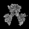 | |||||||||
|---|---|---|---|---|---|---|---|---|---|---|
| Title | CryoEM structure of the HCMV Pentamer gH/gL/UL128/UL130/UL131A in complex with THBD and neutralizing fabs MSL-109 and 13H11 | |||||||||
 Map data Map data | Composite map obtained by combining focused, sharpened maps. Map used for model building and refinements. | |||||||||
 Sample Sample |
| |||||||||
| Function / homology |  Function and homology information Function and homology information blood coagulation, common pathway / negative regulation of blood coagulation / apicolateral plasma membrane / serine-type endopeptidase complex / zymogen activation / vacuolar membrane / negative regulation of platelet activation / response to X-ray / negative regulation of fibrinolysis / Common Pathway of Fibrin Clot Formation ...blood coagulation, common pathway / negative regulation of blood coagulation / apicolateral plasma membrane / serine-type endopeptidase complex / zymogen activation / vacuolar membrane / negative regulation of platelet activation / response to X-ray / negative regulation of fibrinolysis / Common Pathway of Fibrin Clot Formation / response to cAMP / host cell endosome membrane / female pregnancy / Cell surface interactions at the vascular wall / transmembrane signaling receptor activity / blood coagulation / signaling receptor activity / host cell Golgi apparatus / response to lipopolysaccharide / entry receptor-mediated virion attachment to host cell / symbiont entry into host cell / fusion of virus membrane with host plasma membrane / external side of plasma membrane / viral envelope / calcium ion binding / host cell plasma membrane / virion membrane / cell surface / proteolysis / extracellular space / membrane / plasma membrane blood coagulation, common pathway / negative regulation of blood coagulation / apicolateral plasma membrane / serine-type endopeptidase complex / zymogen activation / vacuolar membrane / negative regulation of platelet activation / response to X-ray / negative regulation of fibrinolysis / Common Pathway of Fibrin Clot Formation ...blood coagulation, common pathway / negative regulation of blood coagulation / apicolateral plasma membrane / serine-type endopeptidase complex / zymogen activation / vacuolar membrane / negative regulation of platelet activation / response to X-ray / negative regulation of fibrinolysis / Common Pathway of Fibrin Clot Formation / response to cAMP / host cell endosome membrane / female pregnancy / Cell surface interactions at the vascular wall / transmembrane signaling receptor activity / blood coagulation / signaling receptor activity / host cell Golgi apparatus / response to lipopolysaccharide / entry receptor-mediated virion attachment to host cell / symbiont entry into host cell / fusion of virus membrane with host plasma membrane / external side of plasma membrane / viral envelope / calcium ion binding / host cell plasma membrane / virion membrane / cell surface / proteolysis / extracellular space / membrane / plasma membraneSimilarity search - Function | |||||||||
| Biological species |  Homo sapiens (human) / Homo sapiens (human) /   Human betaherpesvirus 5 Human betaherpesvirus 5 | |||||||||
| Method | single particle reconstruction / cryo EM / Resolution: 3.3 Å | |||||||||
 Authors Authors | Kschonsak M / Johnson MC / Schelling R / Green EM / Rouge L / Ho H / Patel N / Kilic C / Kraft E / Arthur CP ...Kschonsak M / Johnson MC / Schelling R / Green EM / Rouge L / Ho H / Patel N / Kilic C / Kraft E / Arthur CP / Rohou AL / Comps-Agrar L / Martinez-Martin N / Perez L / Payandeh J / Ciferri C | |||||||||
| Funding support | 1 items
| |||||||||
 Citation Citation | Journal: Sci Adv / Year: 2022 Title: Structural basis for HCMV Pentamer receptor recognition and antibody neutralization. Authors: Marc Kschonsak / Matthew C Johnson / Rachel Schelling / Evan M Green / Lionel Rougé / Hoangdung Ho / Nidhi Patel / Cem Kilic / Edward Kraft / Christopher P Arthur / Alexis L Rohou / ...Authors: Marc Kschonsak / Matthew C Johnson / Rachel Schelling / Evan M Green / Lionel Rougé / Hoangdung Ho / Nidhi Patel / Cem Kilic / Edward Kraft / Christopher P Arthur / Alexis L Rohou / Laetitia Comps-Agrar / Nadia Martinez-Martin / Laurent Perez / Jian Payandeh / Claudio Ciferri /   Abstract: Human cytomegalovirus (HCMV) represents the viral leading cause of congenital birth defects and uses the gH/gL/UL128-130-131A complex (Pentamer) to enter different cell types, including epithelial ...Human cytomegalovirus (HCMV) represents the viral leading cause of congenital birth defects and uses the gH/gL/UL128-130-131A complex (Pentamer) to enter different cell types, including epithelial and endothelial cells. Upon infection, Pentamer elicits the most potent neutralizing response against HCMV, representing a key vaccine candidate. Despite its relevance, the structural basis for Pentamer receptor recognition and antibody neutralization is largely unknown. Here, we determine the structures of Pentamer bound to neuropilin 2 (NRP2) and a set of potent neutralizing antibodies against HCMV. Moreover, we identify thrombomodulin (THBD) as a functional HCMV receptor and determine the structures of the Pentamer-THBD complex. Unexpectedly, both NRP2 and THBD also promote dimerization of Pentamer. Our results provide a framework for understanding HCMV receptor engagement, cell entry, antibody neutralization, and outline strategies for antiviral therapies against HCMV. | |||||||||
| History |
|
- Structure visualization
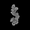
| Supplemental images |
|---|

- Downloads & links
-EMDB archive
| Map data | emd_25686.map.gz | 13 MB | EMDB map data format | |
|---|---|---|---|---|
| Header (meta data) | emd-25686-v30.xmlemd-25686.xml | 59.9 KB 59.9 KB | Display Display | EMDB header |
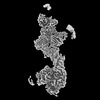
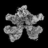
| Images |  emd_25686.png emd_25686.png | 83.3 KB | ||
| Others | emd_25686_additional_1.map.gzemd_25686_additional_10.map.gzemd_25686_additional_11.map.gzemd_25686_additional_12.map.gzemd_25686_additional_13.map.gzemd_25686_additional_14.map.gzemd_25686_additional_15.map.gzemd_25686_additional_16.map.gzemd_25686_additional_2.map.gzemd_25686_additional_3.map.gzemd_25686_additional_4.map.gzemd_25686_additional_5.map.gzemd_25686_additional_6.map.gzemd_25686_additional_7.map.gzemd_25686_additional_8.map.gzemd_25686_additional_9.map.gz | 226.2 MB 165.7 MB 165.7 MB 8.2 MB 226.4 MB 165.7 MB 165.7 MB 12.3 MB 165.5 MB 13.7 MB 165.6 MB 226.3 MB 165.6 MB 165.7 MB 8.5 MB 226.3 MB | ||
| Archive directory |  http://ftp.pdbj.org/pub/emdb/structures/EMD-25686ftp://ftp.pdbj.org/pub/emdb/structures/EMD-25686 http://ftp.pdbj.org/pub/emdb/structures/EMD-25686ftp://ftp.pdbj.org/pub/emdb/structures/EMD-25686 | HTTPS FTP |
-Related structure data
| Related structure data | 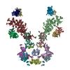 7t4rMC 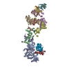 7t4qC 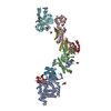 7t4sC M: atomic model generated by this map C: citing same article ( |
|---|---|
| Similar structure data |
-Links
| EMDB pages | EMDB (EBI/PDBe) / EMDataResource |
|---|---|
| Related items in Molecule of the Month |


-Map
| File | Download / File: emd_25686.map.gz / Format: CCP4 / Size: 244.1 MB / Type: IMAGE STORED AS FLOATING POINT NUMBER (4 BYTES) | ||||||||||||||||||||
|---|---|---|---|---|---|---|---|---|---|---|---|---|---|---|---|---|---|---|---|---|---|
| Annotation | Composite map obtained by combining focused, sharpened maps. Map used for model building and refinements. | ||||||||||||||||||||
| Voxel size | X=Y=Z: 1.0726 Å | ||||||||||||||||||||
| Density |
| ||||||||||||||||||||
| Symmetry | Space group: 1 | ||||||||||||||||||||
| Details | EMDB XML:
|
-Supplemental data
+Additional map: Non-sharpened, full map of overall map (not focused).
 Z
Z Y
Y X
X


+Additional map: Half map 1 of focused gH-gL (A) region.


+Additional map: Half map 2 of focused gH-gL (A) region.


+Additional map: Density modified map of focused gH-gL (A) map...


+Additional map: Non-sharpened, full map of focused gH-gL (B) region.


+Additional map: Half map 1 of focused gH-gL (B) region.


+Additional map: Half map 2 of focused gH-gL (B) region.


+Additional map: Density modified map of overall map (not focused)...


+Additional map: Half map 1 of overall map (not focused).


+Additional map: Density modified map of focused THBD-ULs map used...


+Additional map: Half map 2 of overall map (not focused).


+Additional map: Non-sharpened, full map of focused THBD-ULs region.


+Additional map: Half map 1 of focused THBD-ULs region.


+Additional map: Half map 2 of focused THBD-ULs region.


+Additional map: Density modified map of focused gH-gL (B) map...


+Additional map: Non-sharpened, full map of focused gH-gL (A) region.


- Sample components
+Entire : Complex of 2x HCMV Pentamer gH, gL, UL128, UL130, UL131A bound to...
+Supramolecule #1: Complex of 2x HCMV Pentamer gH, gL, UL128, UL130, UL131A bound to...
+Macromolecule #1: Thrombomodulin
+Macromolecule #2: Envelope glycoprotein H
+Macromolecule #3: Envelope glycoprotein L
+Macromolecule #4: Envelope protein UL128
+Macromolecule #5: Envelope glycoprotein UL130
+Macromolecule #6: Envelope protein UL131A
+Macromolecule #7: Fab 13H11 heavy chain

+Macromolecule #8: Fab 13H11 light chain
+Macromolecule #9: Fab MSL-109 light chain
+Macromolecule #10: Fab MSL-109 heavy chain
+Macromolecule #11: 2-acetamido-2-deoxy-beta-D-glucopyranose

-Experimental details
-Structure determination
| Method | cryo EM |
|---|---|
 Processing Processing | single particle reconstruction |
| Aggregation state | particle |
-Sample preparation
| Concentration | 0.6 mg/mL | |||||||||
|---|---|---|---|---|---|---|---|---|---|---|
| Buffer | pH: 7.5 Component:
| |||||||||
| Grid | Model: UltrAuFoil R0.6/1 / Material: GOLD / Mesh: 300 Details: The grid was incubated with a thiol reactive self-assembling reaction mixture of 4mM monothiolalkane(C11)PEG6-OH (11-mercaptoundecyl) hexaethyleneglycol, (SPT-0011P6, SensoPath Technologies, ...Details: The grid was incubated with a thiol reactive self-assembling reaction mixture of 4mM monothiolalkane(C11)PEG6-OH (11-mercaptoundecyl) hexaethyleneglycol, (SPT-0011P6, SensoPath Technologies, Inc., Bozeman, MT)[23]. Grids were incubated with this self-assembled, monolayer (SAM) solution for 24 hours. Prior to grid freezing, grids were removed from the SAM solution and rinsed with EtOH. | |||||||||
| Vitrification | Cryogen name: ETHANE / Chamber humidity: 100 % / Chamber temperature: 277.15 K / Instrument: LEICA EM GP / Details: blot for 3.5s before plunging. |
- Electron microscopy
| Microscope | FEI TITAN KRIOS |
|---|---|
| Electron beam | Acceleration voltage: 300 kV / Electron source: FIELD EMISSION GUN |
| Electron optics | Illumination mode: FLOOD BEAM / Imaging mode: BRIGHT FIELDBright-field microscopy / Nominal defocus max: 1.5 µm / Nominal defocus min: 0.5 µm / Nominal magnification: 105000 |
| Sample stage | Specimen holder model: FEI TITAN KRIOS AUTOGRID HOLDER / Cooling holder cryogen: NITROGEN |
| Image recording | Film or detector model: GATAN K3 BIOQUANTUM (6k x 4k) / Number grids imaged: 1 / Number real images: 10926 / Average electron dose: 64.0 e/Å2 |
| Experimental equipment |  Model: Titan Krios / Image courtesy: FEI Company |
-Image processing
| Particle selection | Number selected: 10680163 |
|---|---|
| CTF correction | Software - Name: CTFFIND (ver. 4.1.13) |
| Startup model | Type of model: OTHER / Details: ab-initio using C2 symmetry in cisTEM |
| Initial angle assignment | Type: MAXIMUM LIKELIHOOD / Software - Name: RELION (ver. 3.1) |
| Final 3D classification | Number classes: 27 / Software - Name: RELION (ver. 3.1) |
| Final angle assignment | Type: OTHER Details: Manual refine in cisTEM. Map was divided in 3 sections to refine map with focused refinements. |
| Final reconstruction | Applied symmetry - Point group: C1 (asymmetric) / Resolution.type: BY AUTHOR / Resolution: 3.3 Å / Resolution method: FSC 0.143 CUT-OFF / Software - Name: cisTEM (ver. 1.02) Details: A composite map was generated from the three individual focused 3D maps. Number images used: 504492 |
| Details | Movie frames were corrected for motion and aligned. Images with a CTF fit resolution of 5.0 A or better were selected for particle picking. |
