 Movie
Movie Controller

[English] 日本語
 Yorodumi
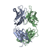
- PDB-7st8: Crystal structure of 7H2.2 Fab in complex with SAS1B C-terminal region -
+ Open data

- Basic information
| Entry | Database: PDB / ID: 7st8 | ||||||
|---|---|---|---|---|---|---|---|
| Title | Crystal structure of 7H2.2 Fab in complex with SAS1B C-terminal region | ||||||
 Components Components |
| ||||||
 Keywords Keywords |  IMMUNE SYSTEM / Cancer / antibody / oocyte antigen IMMUNE SYSTEM / Cancer / antibody / oocyte antigen | ||||||
| Function / homology |  Function and homology information Function and homology informationglutamic-type peptidase activity / negative regulation of binding of sperm to zona pellucida / aspartic-type peptidase activity / prevention of polyspermy / cortical granule / positive regulation of protein processing / fertilization / Hydrolases; Acting on peptide bonds (peptidases) / metalloendopeptidase activity / cell adhesion ...glutamic-type peptidase activity / negative regulation of binding of sperm to zona pellucida / aspartic-type peptidase activity / prevention of polyspermy / cortical granule / positive regulation of protein processing / fertilization / Hydrolases; Acting on peptide bonds (peptidases) / metalloendopeptidase activity / cell adhesion / zinc ion binding / plasma membrane / cytoplasmSimilarity search - Function | ||||||
| Biological species |  Mus musculus (house mouse) Mus musculus (house mouse) Homo sapiens (human) Homo sapiens (human) | ||||||
| Method | X-RAY DIFFRACTION / MOLECULAR REPLACEMENT / Resolution: 2.75 Å | ||||||
 Authors Authors | Legg, M.S.G. / Evans, S.V. | ||||||
| Funding support |  Canada, 1items Canada, 1items
| ||||||
 Citation Citation | Journal: Acta Crystallogr.,Sect.D / Year: 2022 Title: Monoclonal antibody 7H2.2 binds the C-terminus of the cancer-oocyte antigen SAS1B through the hydrophilic face of a conserved amphipathic helix corresponding to one of only two regions predicted to be ordered Authors: Legg, M.S.G. / Gagnon, S.M.L. / Powell, C.J. / Boulanger, M.J. / Li, A.J.J. / Evans, S.V. | ||||||
| History |
|
- Structure visualization
| Structure viewer | Molecule: MolmilJmol/JSmol |
|---|
- Downloads & links
-Download
| PDBx/mmCIF format | 7st8.cif.gz | 104.4 KB | Display | PDBx/mmCIF format |
|---|---|---|---|---|
| PDB format | pdb7st8.ent.gz | 74.7 KB | Display | PDB format |
| PDBx/mmJSON format | 7st8.json.gz | Tree view | PDBx/mmJSON format | |
| Others |  Other downloads Other downloads |
-Validation report
| Arichive directory | https://data.pdbj.org/pub/pdb/validation_reports/st/7st8ftp://data.pdbj.org/pub/pdb/validation_reports/st/7st8 | HTTPS FTP |
|---|
-Related structure data

-Links
 PDBj
PDBj


- Assembly
| Deposited unit | 
| ||||||||
|---|---|---|---|---|---|---|---|---|---|
| 1 |
| ||||||||
| Unit cell |
|
-Components
| #1: Antibody | Mass: 23838.760 Da / Num. of mol.: 1 Source method: isolated from a genetically manipulated source Source: (gene. exp.) Mus musculus (house mouse) / Production host: Mus musculus (house mouse) |
|---|---|
| #2: Antibody | Mass: 23852.354 Da / Num. of mol.: 1 Source method: isolated from a genetically manipulated source Source: (gene. exp.) Mus musculus (house mouse) / Production host: Mus musculus (house mouse) |
| #3: Protein | Mass: 17208.973 Da / Num. of mol.: 1 / Fragment: C-terminus Source method: isolated from a genetically manipulated source Source: (gene. exp.) Homo sapiens (human) / Gene: ASTL / Production host:  Escherichia coli (E. coli) Escherichia coli (E. coli)References: UniProt: Q6HA08, Hydrolases; Acting on peptide bonds (peptidases) |
| #4: Water | ChemComp-HOH / Water Mass: 18.015 Da / Num. of mol.: 26 / Source method: isolated from a natural source / Formula: H2O Mass: 18.015 Da / Num. of mol.: 26 / Source method: isolated from a natural source / Formula: H2O |
-Experimental details
-Experiment
| Experiment | Method: X-RAY DIFFRACTION / Number of used crystals: 1 |
|---|
- Sample preparation
| Crystal | Density Matthews: 1.93 Å3/Da / Density % sol: 36.42 % |
|---|---|
| Crystal grow | Temperature: 291.15 K / Method: vapor diffusion, sitting drop / Details: 0.1 M CHES, pH 9.5, 20 % (w/v) PEG 8000 |
-Data collection
| Diffraction | Mean temperature: 100 K / Serial crystal experiment: N | |||||||||||||||||||||||||||||||||||||||||||||||||||||||||||||||||||||||||||||||||||||||||||||||||||
|---|---|---|---|---|---|---|---|---|---|---|---|---|---|---|---|---|---|---|---|---|---|---|---|---|---|---|---|---|---|---|---|---|---|---|---|---|---|---|---|---|---|---|---|---|---|---|---|---|---|---|---|---|---|---|---|---|---|---|---|---|---|---|---|---|---|---|---|---|---|---|---|---|---|---|---|---|---|---|---|---|---|---|---|---|---|---|---|---|---|---|---|---|---|---|---|---|---|---|---|---|
| Diffraction source | Source: ROTATING ANODE / Type: RIGAKU MICROMAX-007 HF / Wavelength: 1.5418 Å | |||||||||||||||||||||||||||||||||||||||||||||||||||||||||||||||||||||||||||||||||||||||||||||||||||
| Detector | Type: DECTRIS PILATUS 200K / Detector: PIXEL / Date: Dec 18, 2018 | |||||||||||||||||||||||||||||||||||||||||||||||||||||||||||||||||||||||||||||||||||||||||||||||||||
| Radiation | Protocol: SINGLE WAVELENGTH / Monochromatic (M) / Laue (L): M / Scattering type: x-ray | |||||||||||||||||||||||||||||||||||||||||||||||||||||||||||||||||||||||||||||||||||||||||||||||||||
| Radiation wavelength | Wavelength: 1.5418 Å / Relative weight: 1 | |||||||||||||||||||||||||||||||||||||||||||||||||||||||||||||||||||||||||||||||||||||||||||||||||||
| Reflection | Resolution: 2.75→40 Å / Num. obs: 13315 / % possible obs: 96.9 % / Redundancy: 9.7 % / Rmerge(I) obs: 0.161 / Rpim(I) all: 0.049 / Rrim(I) all: 0.169 / Χ2: 1.146 / Net I/σ(I): 16.9 / Num. measured all: 129654 | |||||||||||||||||||||||||||||||||||||||||||||||||||||||||||||||||||||||||||||||||||||||||||||||||||
| Reflection shell | Diffraction-ID: 1
|
- Processing
| Software |
| ||||||||||||||||||||||||||||||||||||||||||||||||||||||||||||
|---|---|---|---|---|---|---|---|---|---|---|---|---|---|---|---|---|---|---|---|---|---|---|---|---|---|---|---|---|---|---|---|---|---|---|---|---|---|---|---|---|---|---|---|---|---|---|---|---|---|---|---|---|---|---|---|---|---|---|---|---|---|
| Refinement | Method to determine structure: MOLECULAR REPLACEMENT Starting model: 3U9U Fab heavy chain, and 2XQY Fab light chain Resolution: 2.75→26.41 Å / Cor.coef. Fo:Fc: 0.915 / Cor.coef. Fo:Fc free: 0.88 / SU B: 19.049 / SU ML: 0.36 / Cross valid method: THROUGHOUT / σ(F): 0 / ESU R Free: 0.415 / Stereochemistry target values: MAXIMUM LIKELIHOOD Details: HYDROGENS HAVE BEEN ADDED IN THE RIDING POSITIONS U VALUES : REFINED INDIVIDUALLY
| ||||||||||||||||||||||||||||||||||||||||||||||||||||||||||||
| Solvent computation | Ion probe radii: 0.8 Å / Shrinkage radii: 0.8 Å / VDW probe radii: 1.2 Å / Solvent model: MASK | ||||||||||||||||||||||||||||||||||||||||||||||||||||||||||||
| Displacement parameters | Biso max: 104.09 Å2 / Biso mean: 40.324 Å2 / Biso min: 0.5 Å2
| ||||||||||||||||||||||||||||||||||||||||||||||||||||||||||||
| Refinement step | Cycle: final / Resolution: 2.75→26.41 Å
| ||||||||||||||||||||||||||||||||||||||||||||||||||||||||||||
| Refine LS restraints |
| ||||||||||||||||||||||||||||||||||||||||||||||||||||||||||||
| LS refinement shell | Resolution: 2.75→2.821 Å / Rfactor Rfree error: 0 / Total num. of bins used: 20
|
