 Movie
Movie Controller

[English] 日本語
 Yorodumi
- PDB-7sql: Crystal structure of human uridine-cytidine kinase 2 complexed wi... -
+ Open data

- Basic information
| Entry | Database: PDB / ID: 7sql | ||||||
|---|---|---|---|---|---|---|---|
| Title | Crystal structure of human uridine-cytidine kinase 2 complexed with a weak small molecule inhibitor | ||||||
 Components Components | Uridine-cytidine kinase 2 | ||||||
 Keywords Keywords | Transferase/Inhibitor /  Kinase / Uridine kinase / Cytidine kinase / Inhibitor / transferase / Transferase-Inhibitor complex Kinase / Uridine kinase / Cytidine kinase / Inhibitor / transferase / Transferase-Inhibitor complex | ||||||
| Function / homology |  Function and homology information Function and homology informationuridine/cytidine kinase / CTP salvage / ribosylnicotinamide kinase activity / uridine kinase activity / Pyrimidine salvage / cytidine kinase activity / UMP salvage / phosphorylation / ATP binding / identical protein binding ...uridine/cytidine kinase / CTP salvage / ribosylnicotinamide kinase activity / uridine kinase activity / Pyrimidine salvage / cytidine kinase activity / UMP salvage / phosphorylation / ATP binding / identical protein binding / cytosol / cytoplasmSimilarity search - Function | ||||||
| Biological species |  Homo sapiens (human) Homo sapiens (human) | ||||||
| Method | X-RAY DIFFRACTION / SYNCHROTRON / MOLECULAR REPLACEMENT / molecular replacement / Resolution: 2.4 Å | ||||||
 Authors Authors | Mashayekh, S. / Stunkard, L.M. / Kienle, M. / Mathews, I.I. / Khosla, C. | ||||||
| Funding support | 1items
| ||||||
 Citation Citation | Journal: Biochemistry / Year: 2022 Title: Structure-Based Prototyping of Allosteric Inhibitors of Human Uridine/Cytidine Kinase 2 (UCK2). Authors: Mashayekh, S. / Stunkard, L.M. / Kienle, M. / Mathews, I.I. / Khosla, C. | ||||||
| History |
|
- Structure visualization
| Structure viewer | Molecule: MolmilJmol/JSmol |
|---|
- Downloads & links
-Download
| PDBx/mmCIF format | 7sql.cif.gz | 192.4 KB | Display | PDBx/mmCIF format |
|---|---|---|---|---|
| PDB format | pdb7sql.ent.gz | 153.3 KB | Display | PDB format |
| PDBx/mmJSON format | 7sql.json.gz | Tree view | PDBx/mmJSON format | |
| Others |  Other downloads Other downloads |
-Validation report
| Arichive directory | https://data.pdbj.org/pub/pdb/validation_reports/sq/7sqlftp://data.pdbj.org/pub/pdb/validation_reports/sq/7sql | HTTPS FTP |
|---|
-Related structure data
| Related structure data | 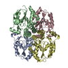 1ufqS S: Starting model for refinement |
|---|---|
| Similar structure data |
-Links
 PDBj
PDBj- Assembly
| Deposited unit | 
| ||||||||
|---|---|---|---|---|---|---|---|---|---|
| 1 |
| ||||||||
| Unit cell |
|
-Components
-Protein , 1 types, 4 molecules ABCD
| #1: Protein | Mass: 28108.830 Da / Num. of mol.: 4 Source method: isolated from a genetically manipulated source Source: (gene. exp.) Homo sapiens (human) / Gene: UCK2, UMPK / Production host:  Escherichia coli BL21(DE3) (bacteria) / References: UniProt: Q9BZX2, uridine/cytidine kinase Escherichia coli BL21(DE3) (bacteria) / References: UniProt: Q9BZX2, uridine/cytidine kinase |
|---|
-Non-polymers , 6 types, 246 molecules 


| #2: Chemical | Polyethylene glycol Mass: 150.173 Da / Num. of mol.: 2 / Source method: isolated from a natural source / Formula: C6H14O4 Mass: 150.173 Da / Num. of mol.: 2 / Source method: isolated from a natural source / Formula: C6H14O4#3: Chemical | ChemComp-GOL / Glycerol Mass: 92.094 Da / Num. of mol.: 4 / Source method: obtained synthetically / Formula: C3H8O3 Mass: 92.094 Da / Num. of mol.: 4 / Source method: obtained synthetically / Formula: C3H8O3#4: Chemical | Polyethylene glycol Mass: 194.226 Da / Num. of mol.: 2 / Source method: isolated from a natural source / Formula: C8H18O5 / Comment: precipitant*YM Mass: 194.226 Da / Num. of mol.: 2 / Source method: isolated from a natural source / Formula: C8H18O5 / Comment: precipitant*YM#5: Chemical | Diethylene glycol Mass: 106.120 Da / Num. of mol.: 3 / Source method: isolated from a natural source / Formula: C4H10O3 Mass: 106.120 Da / Num. of mol.: 3 / Source method: isolated from a natural source / Formula: C4H10O3#6: Chemical |  Mass: 474.306 Da / Num. of mol.: 2 / Source method: obtained synthetically / Formula: C19H13BrFN5O2S / Feature type: SUBJECT OF INVESTIGATION Mass: 474.306 Da / Num. of mol.: 2 / Source method: obtained synthetically / Formula: C19H13BrFN5O2S / Feature type: SUBJECT OF INVESTIGATION#7: Water | ChemComp-HOH / | WaterMass: 18.015 Da / Num. of mol.: 233 / Source method: isolated from a natural source / Formula: H2O |
|---|
-Details
| Has ligand of interest | Y |
|---|
-Experimental details
-Experiment
| Experiment | Method: X-RAY DIFFRACTION / Number of used crystals: 1 |
|---|
- Sample preparation
| Crystal | Density Matthews: 2.76 Å3/Da / Density % sol: 55.51 % |
|---|---|
| Crystal grow | Temperature: 293 K / Method: vapor diffusion, hanging drop / pH: 6.9 Details: 4% glycerol, 100 mM HEPES pH 6.9, 8% PEG 3350, and 30% PEG 400 Temp details: room temperature |
-Data collection
| Diffraction | Mean temperature: 100 K Ambient temp details: liquid nitrogen stream at room temperature Serial crystal experiment: N | ||||||||||||||||||||||||||||||
|---|---|---|---|---|---|---|---|---|---|---|---|---|---|---|---|---|---|---|---|---|---|---|---|---|---|---|---|---|---|---|---|
| Diffraction source | Source: SYNCHROTRON / Site: SSRL  / Beamline: BL12-2 / Wavelength: 0.9795 Å / Beamline: BL12-2 / Wavelength: 0.9795 Å | ||||||||||||||||||||||||||||||
| Detector | Type: DECTRIS PILATUS 6M / Detector: PIXEL / Date: Nov 12, 2020 Details: Flat Si Rh coated M0, Kirkpatrick-Baez flat bent Si M1 & M2 | ||||||||||||||||||||||||||||||
| Radiation | Monochromator: Si (111) / Protocol: SINGLE WAVELENGTH / Monochromatic (M) / Laue (L): M / Scattering type: x-ray | ||||||||||||||||||||||||||||||
| Radiation wavelength | Wavelength: 0.9795 Å / Relative weight: 1 | ||||||||||||||||||||||||||||||
| Reflection | Resolution: 2.4→39.7 Å / Num. obs: 49319 / % possible obs: 99.6 % / Redundancy: 11.9 % / CC1/2: 0.995 / Rmerge(I) obs: 0.233 / Rpim(I) all: 0.073 / Rrim(I) all: 0.245 / Net I/σ(I): 11.4 | ||||||||||||||||||||||||||||||
| Reflection shell | Diffraction-ID: 1
|
-Phasing
| Phasing | Method: molecular replacement | ||||||
|---|---|---|---|---|---|---|---|
| Phasing MR | Model details: Phaser MODE: MR_AUTO
|
- Processing
| Software |
| ||||||||||||||||||||||||||||||||||||||||||||||||||||||||||||
|---|---|---|---|---|---|---|---|---|---|---|---|---|---|---|---|---|---|---|---|---|---|---|---|---|---|---|---|---|---|---|---|---|---|---|---|---|---|---|---|---|---|---|---|---|---|---|---|---|---|---|---|---|---|---|---|---|---|---|---|---|---|
| Refinement | Method to determine structure: MOLECULAR REPLACEMENT Starting model: 1UFQ Resolution: 2.4→39.7 Å / Cor.coef. Fo:Fc: 0.947 / Cor.coef. Fo:Fc free: 0.916 / WRfactor Rfree: 0.238 / WRfactor Rwork: 0.1829 / FOM work R set: 0.8064 / SU B: 8.534 / SU ML: 0.194 / SU R Cruickshank DPI: 0.3172 / SU Rfree: 0.2488 / Cross valid method: THROUGHOUT / σ(F): 0 / ESU R: 0.317 / ESU R Free: 0.249 / Stereochemistry target values: MAXIMUM LIKELIHOOD Details: HYDROGENS HAVE BEEN ADDED IN THE RIDING POSITIONS U VALUES : REFINED INDIVIDUALLY
| ||||||||||||||||||||||||||||||||||||||||||||||||||||||||||||
| Solvent computation | Ion probe radii: 0.8 Å / Shrinkage radii: 0.8 Å / VDW probe radii: 1.2 Å / Solvent model: MASK | ||||||||||||||||||||||||||||||||||||||||||||||||||||||||||||
| Displacement parameters | Biso max: 138.21 Å2 / Biso mean: 48.388 Å2 / Biso min: 4.82 Å2
| ||||||||||||||||||||||||||||||||||||||||||||||||||||||||||||
| Refinement step | Cycle: final / Resolution: 2.4→39.7 Å
| ||||||||||||||||||||||||||||||||||||||||||||||||||||||||||||
| Refine LS restraints |
| ||||||||||||||||||||||||||||||||||||||||||||||||||||||||||||
| LS refinement shell | Resolution: 2.4→2.462 Å / Rfactor Rfree error: 0
|
