 Movie
Movie Controller

[English] 日本語
 Yorodumi
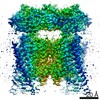
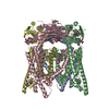
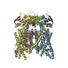
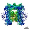
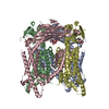
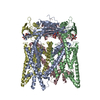
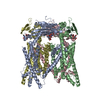
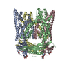
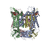
- PDB-7sq7: Cryo-EM structure of mouse PI(3,5)P2-bound TRPML1 channel at 2.41... -
+ Open data

- Basic information
| Entry | Database: PDB / ID: 7sq7 | |||||||||
|---|---|---|---|---|---|---|---|---|---|---|
| Title | Cryo-EM structure of mouse PI(3,5)P2-bound TRPML1 channel at 2.41 Angstrom resolution | |||||||||
 Components Components | Mucolipin-1 MCOLN1 MCOLN1 | |||||||||
 Keywords Keywords | MEMBRANE PROTEIN / ion channel | |||||||||
| Function / homology |  Function and homology information Function and homology informationTransferrin endocytosis and recycling / positive regulation of lysosome organization / calcium ion export / intracellularly phosphatidylinositol-3,5-bisphosphate-gated monatomic cation channel activity / phagosome maturation / NAADP-sensitive calcium-release channel activity / cellular response to pH / ligand-gated calcium channel activity / TRP channels / endosomal transport ...Transferrin endocytosis and recycling / positive regulation of lysosome organization / calcium ion export / intracellularly phosphatidylinositol-3,5-bisphosphate-gated monatomic cation channel activity / phagosome maturation / NAADP-sensitive calcium-release channel activity / cellular response to pH / ligand-gated calcium channel activity / TRP channels / endosomal transport / phagocytic cup / intracellular vesicle / autophagosome maturation / monoatomic cation transmembrane transport / localization / release of sequestered calcium ion into cytosol / monoatomic cation channel activity / cellular response to calcium ion / cell projection / calcium ion transmembrane transport / phagocytic vesicle membrane / late endosome / late endosome membrane / protein homotetramerization / adaptive immune response / lysosome / receptor complex / lysosomal membrane / intracellular membrane-bounded organelle / lipid binding / Golgi apparatus / nucleoplasm / membrane / identical protein binding / plasma membraneSimilarity search - Function | |||||||||
| Biological species |  Mus musculus (house mouse) Mus musculus (house mouse) | |||||||||
| Method | ELECTRON MICROSCOPY / single particle reconstruction / cryo EM / Resolution: 2.41 Å | |||||||||
 Authors Authors | Gan, N. / Han, Y. / Jiang, Y. | |||||||||
| Funding support |  United States, 2items United States, 2items
| |||||||||
 Citation Citation | Journal: Proc Natl Acad Sci U S A / Year: 2022 Title: Structural mechanism of allosteric activation of TRPML1 by PI(3,5)P and rapamycin. Authors: Ninghai Gan / Yan Han / Weizhong Zeng / Yan Wang / Jing Xue / Youxing Jiang / Abstract: Transient receptor potential mucolipin 1 (TRPML1) is a Ca-permeable, nonselective cation channel ubiquitously expressed in the endolysosomes of mammalian cells and its loss-of-function mutations are ...Transient receptor potential mucolipin 1 (TRPML1) is a Ca-permeable, nonselective cation channel ubiquitously expressed in the endolysosomes of mammalian cells and its loss-of-function mutations are the direct cause of type IV mucolipidosis (MLIV), an autosomal recessive lysosomal storage disease. TRPML1 is a ligand-gated channel that can be activated by phosphatidylinositol 3,5-bisphosphate [PI(3,5)P] as well as some synthetic small-molecule agonists. Recently, rapamycin has also been shown to directly bind and activate TRPML1. Interestingly, both PI(3,5)P and rapamycin have low efficacy in channel activation individually but together they work cooperatively and activate the channel with high potency. To reveal the structural basis underlying the synergistic activation of TRPML1 by PI(3,5)P and rapamycin, we determined the high-resolution cryoelectron microscopy (cryo-EM) structures of the mouse TRPML1 channel in various states, including apo closed, PI(3,5)P-bound closed, and PI(3,5)P/temsirolimus (a rapamycin analog)-bound open states. These structures, combined with electrophysiology, elucidate the molecular details of ligand binding and provide structural insight into how the TRPML1 channel integrates two distantly bound ligand stimuli and facilitates channel opening. | |||||||||
| History |
|
- Structure visualization
| Movie |
Movie viewer |
|---|---|
| Structure viewer | Molecule: MolmilJmol/JSmol |
- Downloads & links
-Download
| PDBx/mmCIF format | 7sq7.cif.gz | 337.1 KB | Display | PDBx/mmCIF format |
|---|---|---|---|---|
| PDB format | pdb7sq7.ent.gz | 286.7 KB | Display | PDB format |
| PDBx/mmJSON format | 7sq7.json.gz | Tree view | PDBx/mmJSON format | |
| Others |  Other downloads Other downloads |
-Validation report
| Arichive directory | https://data.pdbj.org/pub/pdb/validation_reports/sq/7sq7ftp://data.pdbj.org/pub/pdb/validation_reports/sq/7sq7 | HTTPS FTP |
|---|
-Related structure data
| Related structure data |  25378MC 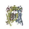 7sq6C  7sq8C 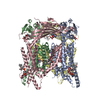 7sq9C M: map data used to model this data C: citing same article ( |
|---|---|
| Similar structure data |


-Links
 PDBj
PDBj- Assembly
| Deposited unit | 
|
|---|---|
| 1 |
|
-Components
| #1: Protein | MCOLN1 / Mucolipidin / Transient receptor potential-mucolipin 1 / TRPML1 Mass: 65573.617 Da / Num. of mol.: 4 Source method: isolated from a genetically manipulated source Source: (gene. exp.) Mus musculus (house mouse) / Gene: Mcoln1, Trpml1 / Production host:  Homo sapiens (human) / References: UniProt: Q99J21 Homo sapiens (human) / References: UniProt: Q99J21#2: Polysaccharide | 2-acetamido-2-deoxy-beta-D-glucopyranose-(1-4)-2-acetamido-2-deoxy-beta-D-glucopyranose / Mass: 424.401 Da / Num. of mol.: 4Source method: isolated from a genetically manipulated source #3: Chemical | ChemComp-EUJ / ( 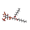  Mass: 746.566 Da / Num. of mol.: 4 / Source method: obtained synthetically / Formula: C25H49O19P3 Mass: 746.566 Da / Num. of mol.: 4 / Source method: obtained synthetically / Formula: C25H49O19P3#4: Chemical | ChemComp-NA / |   Mass: 22.990 Da / Num. of mol.: 1 / Source method: obtained synthetically / Formula: Na Mass: 22.990 Da / Num. of mol.: 1 / Source method: obtained synthetically / Formula: Na#5: Water | ChemComp-HOH / | Water Mass: 18.015 Da / Num. of mol.: 8 / Source method: isolated from a natural source / Formula: H2O Mass: 18.015 Da / Num. of mol.: 8 / Source method: isolated from a natural source / Formula: H2OHas ligand of interest | N | |
|---|
-Experimental details
-Experiment
| Experiment | Method: ELECTRON MICROSCOPY |
|---|---|
| EM experiment | Aggregation state: PARTICLE / 3D reconstruction method: single particle reconstruction |
- Sample preparation
| Component | Name: Mouse Transient receptor potential-mucolipin 1 apo closed state Type: COMPLEX / Entity ID: #1 / Source: RECOMBINANT |
|---|---|
| Source (natural) | Organism: Mus musculus (house mouse) |
| Source (recombinant) | Organism: Homo sapiens (human) |
| Buffer solution | pH: 8 |
| Specimen | Embedding applied: NO / Shadowing applied: NO / Staining applied: NO / Vitrification applied: YES |
| Vitrification | Cryogen name: ETHANE |
- Electron microscopy imaging
| Experimental equipment |  Model: Titan Krios / Image courtesy: FEI Company |
|---|---|
| Microscopy | Model: FEI TITAN KRIOS |
| Electron gun | Electron source: FIELD EMISSION GUN / Accelerating voltage: 300 kV / Illumination mode: FLOOD BEAM |
| Electron lens | Mode: BRIGHT FIELDBright-field microscopy / Nominal defocus max: 2200 nm / Nominal defocus min: 900 nm |
| Image recording | Electron dose: 60 e/Å2 / Film or detector model: GATAN K3 (6k x 4k) |
- Processing
| CTF correction | Type: PHASE FLIPPING ONLY |
|---|---|
| 3D reconstruction | Resolution: 2.41 Å / Resolution method: FSC 0.143 CUT-OFF / Num. of particles: 53514 / Symmetry type: POINT |
