[English] 日本語
Yorodumi
- PDB-7sg5: Structure of PfCSP peptide 21 with antibody CIS43_Var2 -

+
Open data


ID or keywords:

Loading...

-
Basic information

Entry
Database: PDB / ID: 7sg5
TitleStructure of PfCSP peptide 21 with antibody CIS43_Var2
Components
  • (CIS43_Var2 Fab ...) x 2
  • PfCSP peptide 21
KeywordsIMMUNE SYSTEM / Antibody / Plasmodium falciparum
Biological speciesHomo sapiens (human)
synthetic construct (others)
MethodX-RAY DIFFRACTION / SYNCHROTRON / MOLECULAR REPLACEMENT / Resolution: 1.4 Å
AuthorsTripathi, P. / Kwong, P.D.
Funding support United States, 1items
OrganizationGrant numberCountry
National Institutes of Health/National Institute Of Allergy and Infectious Diseases (NIH/NIAID) United States
Citation
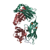
Journal: J Exp Med / Year: 2022
Title: Highly protective antimalarial antibodies via precision library generation and yeast display screening.
Authors: Bailey B Banach / Prabhanshu Tripathi / Lais Da Silva Pereira / Jason Gorman / Thuy Duong Nguyen / Marlon Dillon / Ahmed S Fahad / Patience K Kiyuka / Bharat Madan / Jacy R Wolfe / Brian ...Authors: Bailey B Banach / Prabhanshu Tripathi / Lais Da Silva Pereira / Jason Gorman / Thuy Duong Nguyen / Marlon Dillon / Ahmed S Fahad / Patience K Kiyuka / Bharat Madan / Jacy R Wolfe / Brian Bonilla / Barbara Flynn / Joseph R Francica / Nicholas K Hurlburt / Neville K Kisalu / Tracy Liu / Li Ou / Reda Rawi / Arne Schön / Chen-Hsiang Shen / I-Ting Teng / Baoshan Zhang / Marie Pancera / Azza H Idris / Robert A Seder / Peter D Kwong / Brandon J DeKosky /
Abstract: The monoclonal antibody CIS43 targets the Plasmodium falciparum circumsporozoite protein (PfCSP) and prevents malaria infection in humans for up to 9 mo following a single intravenous administration. ...The monoclonal antibody CIS43 targets the Plasmodium falciparum circumsporozoite protein (PfCSP) and prevents malaria infection in humans for up to 9 mo following a single intravenous administration. To enhance the potency and clinical utility of CIS43, we used iterative site-saturation mutagenesis and DNA shuffling to screen precise gene-variant yeast display libraries for improved PfCSP antigen recognition. We identified several mutations that improved recognition, predominately in framework regions, and combined these to produce a panel of antibody variants. The most improved antibody, CIS43_Var10, had three mutations and showed approximately sixfold enhanced protective potency in vivo compared to CIS43. Co-crystal and cryo-electron microscopy structures of CIS43_Var10 with the peptide epitope or with PfCSP, respectively, revealed functional roles for each of these mutations. The unbiased site-directed mutagenesis and screening pipeline described here represent a powerful approach to enhance protective potency and to enable broader clinical use of antimalarial antibodies.
History
DepositionOct 5, 2021Deposition site: RCSB / Processing site: RCSB
Revision 1.0Jun 22, 2022Provider: repository / Type: Initial release
Revision 1.1Jul 5, 2023Group: Database references / Category: citation / citation_author
Item: _citation.country / _citation.journal_abbrev ..._citation.country / _citation.journal_abbrev / _citation.journal_id_ASTM / _citation.journal_id_CSD / _citation.journal_id_ISSN / _citation.journal_volume / _citation.pdbx_database_id_DOI / _citation.pdbx_database_id_PubMed / _citation.title / _citation.year
Revision 1.2Oct 25, 2023Group: Data collection / Refinement description
Category: chem_comp_atom / chem_comp_bond / pdbx_initial_refinement_model

-
Structure visualization

Structure viewerMolecule:
MolmilJmol/JSmol

Downloads & links

-
Assembly

Deposited unit
H: CIS43_Var2 Fab Heavy chain
L: CIS43_Var2 Fab Light chain
A: PfCSP peptide 21
hetero molecules


Theoretical massNumber of molelcules
Total (without water)50,5899
Polymers50,0173
Non-polymers5726
Water6,702372
1


  • Idetical with deposited unit
  • defined by author&software
  • Evidence: gel filtration
TypeNameSymmetry operationNumber
identity operation1_555x,y,z1
Buried area6280 Å2
ΔGint-85 kcal/mol
Surface area19240 Å2
MethodPISA
Unit cell
Length a, b, c (Å)94.770, 61.137, 75.313
Angle α, β, γ (deg.)90.000, 106.560, 90.000
Int Tables number5
Space group name H-MC121

-
Components

-
Protein/peptide , 1 types, 1 molecules A

#3: Protein/peptide PfCSP peptide 21


Mass: 1562.553 Da / Num. of mol.: 1 / Source method: obtained synthetically / Source: (synth.) synthetic construct (others)

-
Antibody , 2 types, 2 molecules HL

#1: Antibody CIS43_Var2 Fab Heavy chain


Mass: 24281.320 Da / Num. of mol.: 1
Source method: isolated from a genetically manipulated source
Source: (gene. exp.) Homo sapiens (human) / Production host: Homo sapiens (human)
#2: Antibody CIS43_Var2 Fab Light chain


Mass: 24172.723 Da / Num. of mol.: 1
Source method: isolated from a genetically manipulated source
Source: (gene. exp.) Homo sapiens (human) / Production host: Homo sapiens (human)

-
Non-polymers , 3 types, 378 molecules

#4: Chemical
ChemComp-SO4 / SULFATE ION / Sulfate


Mass: 96.063 Da / Num. of mol.: 5 / Source method: obtained synthetically / Formula: SO4
#5: Chemical ChemComp-GOL / GLYCEROL / GLYCERIN / PROPANE-1,2,3-TRIOL / Glycerol


Mass: 92.094 Da / Num. of mol.: 1 / Source method: obtained synthetically / Formula: C3H8O3
#6: Water ChemComp-HOH / water / Water


Mass: 18.015 Da / Num. of mol.: 372 / Source method: isolated from a natural source / Formula: H2O

-
Details

Has ligand of interestN

-
Experimental details

-
Experiment

ExperimentMethod: X-RAY DIFFRACTION / Number of used crystals: 1

-
Sample preparation

CrystalDensity Matthews: 2.09 Å3/Da / Density % sol: 41.17 %
Crystal growTemperature: 298 K / Method: vapor diffusion, hanging drop
Details: 0.2 M ammonium sulfate, 28% w/v polyethylene glycol 4,000

-
Data collection

DiffractionMean temperature: 298 K / Serial crystal experiment: N
Diffraction sourceSource: SYNCHROTRON / Site: APS / Beamline: 22-ID / Wavelength: 1 Å
DetectorType: DECTRIS EIGER X 16M / Detector: PIXEL / Date: Sep 19, 2021
RadiationProtocol: SINGLE WAVELENGTH / Monochromatic (M) / Laue (L): M / Scattering type: x-ray
Radiation wavelengthWavelength: 1 Å / Relative weight: 1
ReflectionResolution: 1.4→50.72 Å / Num. obs: 61516 / % possible obs: 75.52 % / Redundancy: 2.1 % / CC1/2: 0.996 / Rmerge(I) obs: 0.044 / Net I/σ(I): 14.71
Reflection shellResolution: 1.4→1.45 Å / Rmerge(I) obs: 0.349 / Num. unique obs: 4587 / CC1/2: 0.82

-
Processing

Software
NameVersionClassification
PHENIX1.18.2_3874refinement
PDB_EXTRACT3.27data extraction
XDSdata reduction
Aimlessdata scaling
PHASERphasing
RefinementMethod to determine structure: MOLECULAR REPLACEMENT
Starting model: 6B5M
Resolution: 1.4→50.72 Å / SU ML: 0.17 / Cross valid method: THROUGHOUT / σ(F): 1.34 / Phase error: 23.67 / Stereochemistry target values: ML
RfactorNum. reflection% reflection
Rfree0.2176 3047 4.97 %
Rwork0.1851 58277 -
obs0.1867 61324 75.52 %
Solvent computationShrinkage radii: 0.9 Å / VDW probe radii: 1.11 Å / Solvent model: FLAT BULK SOLVENT MODEL
Displacement parametersBiso max: 48.08 Å2 / Biso mean: 19.3857 Å2 / Biso min: 9.18 Å2
Refinement stepCycle: final / Resolution: 1.4→50.72 Å
ProteinNucleic acidLigandSolventTotal
Num. atoms3443 0 31 372 3846
Biso mean--28.36 25.08 -
Num. residues----451
LS refinement shell

Refine-ID: X-RAY DIFFRACTION / Rfactor Rfree error: 0

Resolution (Å)Rfactor RfreeNum. reflection RfreeRfactor RworkNum. reflection Rwork% reflection obs (%)
1.4-1.420.317980.2918187954
1.42-1.450.30941110.2701201258
1.45-1.470.31041220.2501226465
1.47-1.50.29251300.2279248071
1.5-1.530.24451460.2189269677
1.53-1.560.24261440.2144279380
1.56-1.590.28271460.206279581
1.59-1.630.22591350.1971280780
1.63-1.670.22321380.207282080
1.67-1.710.24991350.2045285781
1.71-1.760.25781370.1987279280
1.76-1.820.25731330.1966278079
1.82-1.890.21891280.1956279280
1.89-1.960.22691530.1893281281
1.96-2.050.22171690.1838276980
2.05-2.160.22281560.1806279680
2.16-2.290.24321470.1799276679
2.29-2.470.22721440.186274778
2.47-2.720.23731590.1889263875
2.72-3.110.21231300.1864271177
3.11-3.920.18471540.1674265575

+
About Yorodumi

-
News

-
Feb 9, 2022. New format data for meta-information of EMDB entries

New format data for meta-information of EMDB entries

  • Version 3 of the EMDB header file is now the official format.
  • The previous official version 1.9 will be removed from the archive.

Related info.:EMDB header

External links:wwPDB to switch to version 3 of the EMDB data model

-
Aug 12, 2020. Covid-19 info

Covid-19 info

URL: https://pdbj.org/emnavi/covid19.php

New page: Covid-19 featured information page in EM Navigator.

Related info.:Covid-19 info / Mar 5, 2020. Novel coronavirus structure data

+
Mar 5, 2020. Novel coronavirus structure data

Novel coronavirus structure data

Related info.:Yorodumi Speices / Aug 12, 2020. Covid-19 info

External links:COVID-19 featured content - PDBj / Molecule of the Month (242):Coronavirus Proteases

+
Jan 31, 2019. EMDB accession codes are about to change! (news from PDBe EMDB page)

EMDB accession codes are about to change! (news from PDBe EMDB page)

  • The allocation of 4 digits for EMDB accession codes will soon come to an end. Whilst these codes will remain in use, new EMDB accession codes will include an additional digit and will expand incrementally as the available range of codes is exhausted. The current 4-digit format prefixed with “EMD-” (i.e. EMD-XXXX) will advance to a 5-digit format (i.e. EMD-XXXXX), and so on. It is currently estimated that the 4-digit codes will be depleted around Spring 2019, at which point the 5-digit format will come into force.
  • The EM Navigator/Yorodumi systems omit the EMD- prefix.

Related info.:Q: What is EMD? / ID/Accession-code notation in Yorodumi/EM Navigator

External links:EMDB Accession Codes are Changing Soon! / Contact to PDBj

+
Jul 12, 2017. Major update of PDB

Major update of PDB

  • wwPDB released updated PDB data conforming to the new PDBx/mmCIF dictionary.
  • This is a major update changing the version number from 4 to 5, and with Remediation, in which all the entries are updated.
  • In this update, many items about electron microscopy experimental information are reorganized (e.g. em_software).
  • Now, EM Navigator and Yorodumi are based on the updated data.

External links:wwPDB Remediation / Enriched Model Files Conforming to OneDep Data Standards Now Available in the PDB FTP Archive

-
Yorodumi

Thousand views of thousand structures

  • Yorodumi is a browser for structure data from EMDB, PDB, SASBDB, etc.
  • This page is also the successor to EM Navigator detail page, and also detail information page/front-end page for Omokage search.
  • The word "yorodu" (or yorozu) is an old Japanese word meaning "ten thousand". "mi" (miru) is to see.

Related info.:EMDB / PDB / SASBDB / Comparison of 3 databanks / Yorodumi Search / Aug 31, 2016. New EM Navigator & Yorodumi / Yorodumi Papers / Jmol/JSmol / Function and homology information / Changes in new EM Navigator and Yorodumi

Read more