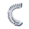
Journal: Nature / Year: 2022 Title: Structural basis for SHOC2 modulation of RAS signalling. Authors: Nicholas P D Liau / Matthew C Johnson / Saeed Izadi / Luca Gerosa / Michal Hammel / John M Bruning / Timothy J Wendorff / Wilson Phung / Sarah G Hymowitz / Jawahar Sudhamsu / Abstract: The RAS-RAF pathway is one of the most commonly dysregulated in human cancers. Despite decades of study, understanding of the molecular mechanisms underlying dimerization and activation of the kinase ...The RAS-RAF pathway is one of the most commonly dysregulated in human cancers. Despite decades of study, understanding of the molecular mechanisms underlying dimerization and activation of the kinase RAF remains limited. Recent structures of inactive RAF monomer and active RAF dimer bound to 14-3-3 have revealed the mechanisms by which 14-3-3 stabilizes both RAF conformations via specific phosphoserine residues. Prior to RAF dimerization, the protein phosphatase 1 catalytic subunit (PP1C) must dephosphorylate the N-terminal phosphoserine (NTpS) of RAF to relieve inhibition by 14-3-3, although PP1C in isolation lacks intrinsic substrate selectivity. SHOC2 is as an essential scaffolding protein that engages both PP1C and RAS to dephosphorylate RAF NTpS, but the structure of SHOC2 and the architecture of the presumptive SHOC2-PP1C-RAS complex remain unknown. Here we present a cryo-electron microscopy structure of the SHOC2-PP1C-MRAS complex to an overall resolution of 3 Å, revealing a tripartite molecular architecture in which a crescent-shaped SHOC2 acts as a cradle and brings together PP1C and MRAS. Our work demonstrates the GTP dependence of multiple RAS isoforms for complex formation, delineates the RAS-isoform preference for complex assembly, and uncovers how the SHOC2 scaffold and RAS collectively drive specificity of PP1C for RAF NTpS. Our data indicate that disease-relevant mutations affect complex assembly, reveal the simultaneous requirement of two RAS molecules for RAF activation, and establish rational avenues for discovery of new classes of inhibitors to target this pathway.
Instrument: FEI VITROBOT MARK IV / Cryogen name: ETHANE / Humidity: 100 % / Chamber temperature: 277 K Details: Used the "perpetually hydrated" method of applying graphene oxide. (Cheung et al., 2018)
-
Electron microscopy imaging
Experimental equipment
Model: Titan Krios / Image courtesy: FEI Company
Microscopy
Model: FEI TITAN KRIOS
Electron gun
Electron source: FIELD EMISSION GUN / Accelerating voltage: 300 kV / Illumination mode: FLOOD BEAM
In the structure databanks used in Yorodumi, some data are registered as the other names, "COVID-19 virus" and "2019-nCoV". Here are the details of the virus and the list of structure data.
Jan 31, 2019. EMDB accession codes are about to change! (news from PDBe EMDB page)
EMDB accession codes are about to change! (news from PDBe EMDB page)
The allocation of 4 digits for EMDB accession codes will soon come to an end. Whilst these codes will remain in use, new EMDB accession codes will include an additional digit and will expand incrementally as the available range of codes is exhausted. The current 4-digit format prefixed with “EMD-” (i.e. EMD-XXXX) will advance to a 5-digit format (i.e. EMD-XXXXX), and so on. It is currently estimated that the 4-digit codes will be depleted around Spring 2019, at which point the 5-digit format will come into force.
The EM Navigator/Yorodumi systems omit the EMD- prefix.
Related info.:Q: What is EMD? / ID/Accession-code notation in Yorodumi/EM Navigator
Yorodumi is a browser for structure data from EMDB, PDB, SASBDB, etc.
This page is also the successor to EM Navigator detail page, and also detail information page/front-end page for Omokage search.
The word "yorodu" (or yorozu) is an old Japanese word meaning "ten thousand". "mi" (miru) is to see.
Related info.:EMDB / PDB / SASBDB / Comparison of 3 databanks / Yorodumi Search / Aug 31, 2016. New EM Navigator & Yorodumi / Yorodumi Papers / Jmol/JSmol / Function and homology information / Changes in new EM Navigator and Yorodumi
 Movie
Movie Controller


 Open data
 Basic information
 Components
 Keywords
 SIGNALING PROTEIN /
  Function and homology information
 Authors
 United States, 1items
  Citation
 Structure visualization
 Downloads & links
 Other downloads
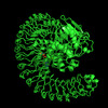
 PDBj
PDBj


 Assembly
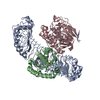


 Mass: 521.208 Da / Num. of mol.: 1 / Source method: obtained synthetically / Formula: C11H18N5O13P3 / Comment: GMP-PCP, energy-carrying molecule analogue*YM
 Mass: 24.305 Da / Num. of mol.: 1 / Source method: obtained synthetically / Formula: Mg
 Mass: 54.938 Da / Num. of mol.: 2 / Source method: obtained synthetically / Formula: Mn
 Sample preparation
 Electron microscopy imaging
 Processing
