 Movie
Movie Controller


+ Open data

- Basic information
| Entry | Database: PDB / ID: 7sc4 | ||||||||||||
|---|---|---|---|---|---|---|---|---|---|---|---|---|---|
| Title | filamin complex-1 | ||||||||||||
 Components Components | Filamin-A/Integrin alpha-IIb light chain, form 2 chimera | ||||||||||||
 Keywords Keywords |  CELL ADHESION / filamin / integrin / inside-out signaling / outside-in signaling CELL ADHESION / filamin / integrin / inside-out signaling / outside-in signaling | ||||||||||||
| Function / homology |  Function and homology information Function and homology informationregulation of membrane repolarization during atrial cardiac muscle cell action potential / regulation of membrane repolarization during cardiac muscle cell action potential / establishment of Sertoli cell barrier / Myb complex / formation of radial glial scaffolds / glycoprotein Ib-IX-V complex / adenylate cyclase-inhibiting dopamine receptor signaling pathway / positive regulation of integrin-mediated signaling pathway / cytoplasmic sequestering of protein / tubulin deacetylation ...regulation of membrane repolarization during atrial cardiac muscle cell action potential / regulation of membrane repolarization during cardiac muscle cell action potential / establishment of Sertoli cell barrier / Myb complex / formation of radial glial scaffolds / glycoprotein Ib-IX-V complex / adenylate cyclase-inhibiting dopamine receptor signaling pathway / positive regulation of integrin-mediated signaling pathway / cytoplasmic sequestering of protein / tubulin deacetylation / actin crosslink formation / blood coagulation, intrinsic pathway / protein localization to bicellular tight junction / OAS antiviral response / positive regulation of actin filament bundle assembly / platelet alpha granule membrane / positive regulation of neuron migration / positive regulation of potassium ion transmembrane transport / Cell-extracellular matrix interactions / fibrinogen binding / early endosome to late endosome transport / apical dendrite / cell-cell junction organization / positive regulation of neural precursor cell proliferation / positive regulation of platelet activation / extracellular matrix binding / protein localization to cell surface / integrin complex / positive regulation of leukocyte migration / wound healing, spreading of cells / negative regulation of transcription by RNA polymerase I / Fc-gamma receptor I complex binding / cell adhesion mediated by integrin / megakaryocyte development / GP1b-IX-V activation signalling / cortical cytoskeleton / p130Cas linkage to MAPK signaling for integrins / receptor clustering / positive regulation of axon regeneration / actin filament bundle / SMAD binding / RHO GTPases activate PAKs / brush border / GRB2:SOS provides linkage to MAPK signaling for Integrins / semaphorin-plexin signaling pathway / cilium assembly / mitotic spindle assembly / ECM proteoglycans / potassium channel regulator activity / epithelial to mesenchymal transition / blood vessel remodeling / Integrin cell surface interactions / axonal growth cone / heart morphogenesis / positive regulation of substrate adhesion-dependent cell spreading / release of sequestered calcium ion into cytosol / regulation of cell migration / Integrin signaling / cell-matrix adhesion / protein kinase C binding / dendritic shaft / Signal transduction by L1 / G protein-coupled receptor binding / actin filament / integrin-mediated signaling pathway / protein localization to plasma membrane / synapse organization / Signaling by high-kinase activity BRAF mutants / RUNX1 regulates genes involved in megakaryocyte differentiation and platelet function / mRNA transcription by RNA polymerase II / MAP2K and MAPK activation / establishment of protein localization / trans-Golgi network / negative regulation of DNA-binding transcription factor activity / negative regulation of protein catabolic process / cell-cell adhesion / cerebral cortex development / platelet aggregation / small GTPase binding / Z disc / kinase binding / positive regulation of protein import into nucleus / Signaling by RAF1 mutants / Signaling by moderate kinase activity BRAF mutants / Paradoxical activation of RAF signaling by kinase inactive BRAF / Signaling downstream of RAS mutants / actin filament binding / Signaling by BRAF and RAF1 fusions / cell-cell junction / actin cytoskeleton / integrin binding / negative regulation of neuron projection development / Platelet degranulation / GTPase binding / perikaryon / actin cytoskeleton organization / postsynapse / angiogenesis / positive regulation of canonical NF-kappaB signal transduction / blood microparticleSimilarity search - Function | ||||||||||||
| Biological species |  Homo sapiens (human) Homo sapiens (human) | ||||||||||||
| Method | X-RAY DIFFRACTION / SYNCHROTRON / MOLECULAR REPLACEMENT / Resolution: 1.85 Å | ||||||||||||
 Authors Authors | Liu, J. / Qin, J. | ||||||||||||
| Funding support |  United States, 3items United States, 3items
| ||||||||||||
 Citation Citation | Journal: Blood / Year: 2023 Title: A mechanism of platelet integrin alpha IIb beta 3 outside-in signaling through a novel integrin alpha IIb subunit-filamin-actin linkage. Authors: Liu, J. / Lu, F. / Ithychanda, S.S. / Apostol, M. / Das, M. / Deshpande, G. / Plow, E.F. / Qin, J. | ||||||||||||
| History |
|
- Structure visualization
| Structure viewer | Molecule: MolmilJmol/JSmol |
|---|
- Downloads & links
-Download
| PDBx/mmCIF format | 7sc4.cif.gz | 94.4 KB | Display | PDBx/mmCIF format |
|---|---|---|---|---|
| PDB format | pdb7sc4.ent.gz | 70.4 KB | Display | PDB format |
| PDBx/mmJSON format | 7sc4.json.gz | Tree view | PDBx/mmJSON format | |
| Others |  Other downloads Other downloads |
-Validation report
| Arichive directory | https://data.pdbj.org/pub/pdb/validation_reports/sc/7sc4ftp://data.pdbj.org/pub/pdb/validation_reports/sc/7sc4 | HTTPS FTP |
|---|
-Related structure data
| Related structure data | 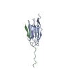 7sftC 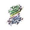 2brqS S: Starting model for refinement C: citing same article ( |
|---|---|
| Similar structure data |
-Links
 PDBj
PDBj


- Assembly
| Deposited unit | 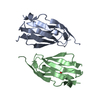
| ||||||||
|---|---|---|---|---|---|---|---|---|---|
| 1 | 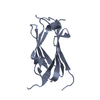
| ||||||||
| 2 | 
| ||||||||
| Unit cell |
|
-Components
| #1: Protein | Mass: 13333.429 Da / Num. of mol.: 2 Source method: isolated from a genetically manipulated source Source: (gene. exp.) Homo sapiens (human) / Gene: FLNA, FLN, FLN1, ITGA2B, GP2B, ITGAB / Production host:  Escherichia phage EcSzw-2 (virus) / References: UniProt: P21333, UniProt: P08514 Escherichia phage EcSzw-2 (virus) / References: UniProt: P21333, UniProt: P08514#2: Chemical | ChemComp-SO4 / | Sulfate  Mass: 96.063 Da / Num. of mol.: 1 / Source method: obtained synthetically / Formula: SO4 Mass: 96.063 Da / Num. of mol.: 1 / Source method: obtained synthetically / Formula: SO4#3: Water | ChemComp-HOH / | Water Mass: 18.015 Da / Num. of mol.: 110 / Source method: isolated from a natural source / Formula: H2O Mass: 18.015 Da / Num. of mol.: 110 / Source method: isolated from a natural source / Formula: H2OHas ligand of interest | N | |
|---|
-Experimental details
-Experiment
| Experiment | Method: X-RAY DIFFRACTION / Number of used crystals: 1 |
|---|
- Sample preparation
| Crystal | Density Matthews: 2.23 Å3/Da / Density % sol: 44.9 % |
|---|---|
| Crystal grow | Temperature: 289.15 K / Method: vapor diffusion, sitting drop / Details: 2 M Ammonium Sulfate, 5% 2-propanol |
-Data collection
| Diffraction | Mean temperature: 100 K / Serial crystal experiment: N |
|---|---|
| Diffraction source | Source: SYNCHROTRON / Site: APS / Beamline: 19-ID / Wavelength: 0.97904 Å |
| Detector | Type: ADSC QUANTUM 315 / Detector: CCD / Date: Apr 14, 2013 |
| Radiation | Monochromator: Si 111 / Protocol: SINGLE WAVELENGTH / Monochromatic (M) / Laue (L): M / Scattering type: x-ray |
| Radiation wavelength | Wavelength: 0.97904 Å / Relative weight: 1 |
| Reflection | Resolution: 1.8→50 Å / Num. obs: 55864 / % possible obs: 99.9 % / Redundancy: 5.3 % / Rmerge(I) obs: 0.11 / Χ2: 1.36 / Net I/σ(I): 20 |
| Reflection shell | Resolution: 2.2→2.24 Å / Redundancy: 4.2 % / Rmerge(I) obs: 0.489 / Mean I/σ(I) obs: 3 / Num. unique obs: 604 / Χ2: 1.12 / % possible all: 98.4 |
- Processing
| Software |
| |||||||||||||||||||||||||||||||||||||||||||||||||||||||||||||||||||||||||||
|---|---|---|---|---|---|---|---|---|---|---|---|---|---|---|---|---|---|---|---|---|---|---|---|---|---|---|---|---|---|---|---|---|---|---|---|---|---|---|---|---|---|---|---|---|---|---|---|---|---|---|---|---|---|---|---|---|---|---|---|---|---|---|---|---|---|---|---|---|---|---|---|---|---|---|---|---|
| Refinement | Method to determine structure: MOLECULAR REPLACEMENT Starting model: 2BRQ Resolution: 1.85→35.97 Å / Cor.coef. Fo:Fc: 0.956 / Cor.coef. Fo:Fc free: 0.927 / SU B: 5.114 / SU ML: 0.095 / Cross valid method: THROUGHOUT / σ(F): 0 / ESU R: 0.139 / ESU R Free: 0.147 / Stereochemistry target values: MAXIMUM LIKELIHOOD Details: HYDROGENS HAVE BEEN USED IF PRESENT IN THE INPUT U VALUES : WITH TLS ADDED
| |||||||||||||||||||||||||||||||||||||||||||||||||||||||||||||||||||||||||||
| Solvent computation | Ion probe radii: 0.8 Å / Shrinkage radii: 0.8 Å / VDW probe radii: 1.2 Å / Solvent model: MASK | |||||||||||||||||||||||||||||||||||||||||||||||||||||||||||||||||||||||||||
| Displacement parameters | Biso max: 88.89 Å2 / Biso mean: 34.845 Å2 / Biso min: 16.79 Å2
| |||||||||||||||||||||||||||||||||||||||||||||||||||||||||||||||||||||||||||
| Refinement step | Cycle: final / Resolution: 1.85→35.97 Å
| |||||||||||||||||||||||||||||||||||||||||||||||||||||||||||||||||||||||||||
| Refine LS restraints |
| |||||||||||||||||||||||||||||||||||||||||||||||||||||||||||||||||||||||||||
| LS refinement shell | Resolution: 1.85→1.896 Å / Rfactor Rfree error: 0
| |||||||||||||||||||||||||||||||||||||||||||||||||||||||||||||||||||||||||||
| Refinement TLS params. | Method: refined / Refine-ID: X-RAY DIFFRACTION
| |||||||||||||||||||||||||||||||||||||||||||||||||||||||||||||||||||||||||||
| Refinement TLS group |
|
