 Movie
Movie Controller

[English] 日本語
 Yorodumi
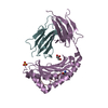
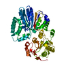
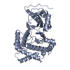
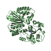
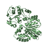
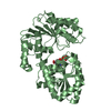
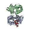
- PDB-7sbh: Crystal structure of the iron superoxide dismutase from Acinetoba... -
+ Open data

- Basic information
| Entry | Database: PDB / ID: 7sbh | ||||||
|---|---|---|---|---|---|---|---|
| Title | Crystal structure of the iron superoxide dismutase from Acinetobacter sp. Ver3 | ||||||
 Components Components | Superoxide dismutase | ||||||
 Keywords Keywords | OXIDOREDUCTASE / metalloenzyme / extremophile / oxidative stress | ||||||
| Function / homology |  Function and homology informationsuperoxide dismutase / superoxide dismutase activity / metal ion binding Function and homology informationsuperoxide dismutase / superoxide dismutase activity / metal ion bindingSimilarity search - Function | ||||||
| Biological species |  Acinetobacter sp. Ver3 (bacteria) Acinetobacter sp. Ver3 (bacteria) | ||||||
| Method | X-RAY DIFFRACTION / SYNCHROTRON / MOLECULAR REPLACEMENT / Resolution: 1.34 Å | ||||||
 Authors Authors | Steimbruch, B.A. / Albanesi, D. / Repizo, G.D. / Lisa, M.N. | ||||||
| Funding support |  Argentina, 1items Argentina, 1items
| ||||||
 Citation Citation | Journal: Sci Rep / Year: 2022 Title: The distinctive roles played by the superoxide dismutases of the extremophile Acinetobacter sp. Ver3. Authors: Steimbruch, B.A. / Sartorio, M.G. / Cortez, N. / Albanesi, D. / Lisa, M.N. / Repizo, G.D. | ||||||
| History |
|
- Structure visualization
| Structure viewer | Molecule: MolmilJmol/JSmol |
|---|
- Downloads & links
-Download
| PDBx/mmCIF format | 7sbh.cif.gz | 125 KB | Display | PDBx/mmCIF format |
|---|---|---|---|---|
| PDB format | pdb7sbh.ent.gz | 78.4 KB | Display | PDB format |
| PDBx/mmJSON format | 7sbh.json.gz | Tree view | PDBx/mmJSON format | |
| Others |  Other downloads Other downloads |
-Validation report
| Arichive directory | https://data.pdbj.org/pub/pdb/validation_reports/sb/7sbhftp://data.pdbj.org/pub/pdb/validation_reports/sb/7sbh | HTTPS FTP |
|---|
-Related structure data
| Related structure data |  1isaS S: Starting model for refinement |
|---|---|
| Similar structure data |


-Links
 PDBj
PDBj
- Assembly
| Deposited unit | 
| ||||||||||||
|---|---|---|---|---|---|---|---|---|---|---|---|---|---|
| 1 | 
| ||||||||||||
| Unit cell |
| ||||||||||||
| Components on special symmetry positions |
|
-Components
| #1: Protein | Mass: 22894.537 Da / Num. of mol.: 1 Source method: isolated from a genetically manipulated source Source: (gene. exp.) Acinetobacter sp. Ver3 (bacteria) / Gene: CL42_08295 / Production host: Escherichia coli (E. coli) / References: UniProt: A0A031LR83, superoxide dismutase |
|---|---|
| #2: Chemical | ChemComp-FMN / Flavin mononucleotide  Mass: 456.344 Da / Num. of mol.: 1 / Source method: obtained synthetically / Formula: C17H21N4O9P / Feature type: SUBJECT OF INVESTIGATION Mass: 456.344 Da / Num. of mol.: 1 / Source method: obtained synthetically / Formula: C17H21N4O9P / Feature type: SUBJECT OF INVESTIGATION |
| #3: Chemical | ChemComp-FE / Iron  Mass: 55.845 Da / Num. of mol.: 1 / Source method: isolated from a natural source / Formula: Fe / Feature type: SUBJECT OF INVESTIGATION Mass: 55.845 Da / Num. of mol.: 1 / Source method: isolated from a natural source / Formula: Fe / Feature type: SUBJECT OF INVESTIGATION |
| #4: Water | ChemComp-HOH / Water Mass: 18.015 Da / Num. of mol.: 352 / Source method: isolated from a natural source / Formula: H2O Mass: 18.015 Da / Num. of mol.: 352 / Source method: isolated from a natural source / Formula: H2O |
| Has ligand of interest | Y |
-Experimental details
-Experiment
| Experiment | Method: X-RAY DIFFRACTION / Number of used crystals: 1 |
|---|
- Sample preparation
| Crystal | Density Matthews: 2.55 Å3/Da / Density % sol: 51.72 % |
|---|---|
| Crystal grow | Temperature: 293 K / Method: vapor diffusion, hanging drop Details: 22 % w/v PEG 8000, 100 mM sodium cacodylate pH 7.5, 200 mM magnesium acetate, 1 mM flavin mononucleotide |
-Data collection
| Diffraction | Mean temperature: 100 K / Serial crystal experiment: N |
|---|---|
| Diffraction source | Source: SYNCHROTRON / Site: Diamond  / Beamline: I04 / Wavelength: 0.9795 Å / Beamline: I04 / Wavelength: 0.9795 Å |
| Detector | Type: DECTRIS EIGER2 XE 16M / Detector: PIXEL / Date: Feb 11, 2020 |
| Radiation | Protocol: SINGLE WAVELENGTH / Monochromatic (M) / Laue (L): M / Scattering type: x-ray |
| Radiation wavelength | Wavelength: 0.9795 Å / Relative weight: 1 |
| Reflection | Resolution: 1.34→28.54 Å / Num. obs: 51999 / % possible obs: 99.7 % / Redundancy: 6.6 % / Biso Wilson estimate: 12.96 Å2 / CC1/2: 0.999 / Rmerge(I) obs: 0.062 / Rpim(I) all: 0.026 / Rrim(I) all: 0.067 / Net I/σ(I): 18.9 |
| Reflection shell | Resolution: 1.34→1.37 Å / Rmerge(I) obs: 0.749 / Mean I/σ(I) obs: 2.4 / Num. unique obs: 2402 / CC1/2: 0.705 / Rpim(I) all: 0.36 / Rrim(I) all: 0.833 |
- Processing
| Software |
| ||||||||||||||||||||||||||||||||||||||||||||||||||||||||||||||||||||||||||||||||||||||||||||||||||||||||||||||||||||||||||||||||||||||||||||
|---|---|---|---|---|---|---|---|---|---|---|---|---|---|---|---|---|---|---|---|---|---|---|---|---|---|---|---|---|---|---|---|---|---|---|---|---|---|---|---|---|---|---|---|---|---|---|---|---|---|---|---|---|---|---|---|---|---|---|---|---|---|---|---|---|---|---|---|---|---|---|---|---|---|---|---|---|---|---|---|---|---|---|---|---|---|---|---|---|---|---|---|---|---|---|---|---|---|---|---|---|---|---|---|---|---|---|---|---|---|---|---|---|---|---|---|---|---|---|---|---|---|---|---|---|---|---|---|---|---|---|---|---|---|---|---|---|---|---|---|---|---|
| Refinement | Method to determine structure: MOLECULAR REPLACEMENT Starting model: 1ISA Resolution: 1.34→27.49 Å / SU ML: 0.1066 / Cross valid method: FREE R-VALUE / σ(F): 1.34 / Phase error: 14.3154 Stereochemistry target values: GeoStd + Monomer Library + CDL v1.2
| ||||||||||||||||||||||||||||||||||||||||||||||||||||||||||||||||||||||||||||||||||||||||||||||||||||||||||||||||||||||||||||||||||||||||||||
| Solvent computation | Shrinkage radii: 0.9 Å / VDW probe radii: 1.11 Å / Solvent model: FLAT BULK SOLVENT MODEL | ||||||||||||||||||||||||||||||||||||||||||||||||||||||||||||||||||||||||||||||||||||||||||||||||||||||||||||||||||||||||||||||||||||||||||||
| Displacement parameters | Biso mean: 17.96 Å2 | ||||||||||||||||||||||||||||||||||||||||||||||||||||||||||||||||||||||||||||||||||||||||||||||||||||||||||||||||||||||||||||||||||||||||||||
| Refinement step | Cycle: LAST / Resolution: 1.34→27.49 Å
| ||||||||||||||||||||||||||||||||||||||||||||||||||||||||||||||||||||||||||||||||||||||||||||||||||||||||||||||||||||||||||||||||||||||||||||
| Refine LS restraints |
| ||||||||||||||||||||||||||||||||||||||||||||||||||||||||||||||||||||||||||||||||||||||||||||||||||||||||||||||||||||||||||||||||||||||||||||
| LS refinement shell |
|
