 Movie
Movie Controller

[English] 日本語
 Yorodumi
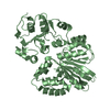
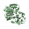
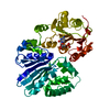
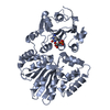
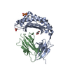
- PDB-4v2u: Apo-structure of alpha2,3-sialyltransferase from Pasteurella dagmatis -
+ Open data

- Basic information
| Entry | Database: PDB / ID: 4v2u | ||||||
|---|---|---|---|---|---|---|---|
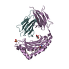
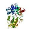
| Title | Apo-structure of alpha2,3-sialyltransferase from Pasteurella dagmatis | ||||||
 Components Components | SIALYLTRANSFERASE | ||||||
 Keywords Keywords | TRANSFERASE | ||||||
| Function / homology |  Function and homology information Function and homology information | ||||||
| Biological species |  PASTEURELLA DAGMATIS (bacteria) PASTEURELLA DAGMATIS (bacteria) | ||||||
| Method | X-RAY DIFFRACTION / SYNCHROTRON / MOLECULAR REPLACEMENT / Resolution: 2.71 Å | ||||||
 Authors Authors | Pavkov-Keller, T. / Schmoelzer, K. / Czabany, T. / Luley-Goedl, C. / Ribitsch, D. / Schwab, H. / Nidetzky, B. / Gruber, K. | ||||||
 Citation Citation | Journal: Chem.Commun.(Camb.) / Year: 2015 Title: Complete Switch from Alpha2,3- to Alpha2,6-Regioselectivity in Pasteurella Dagmatis Beta-D-Galactoside Sialyltransferase by Active-Site Redesign Authors: Schmoelzer, K. / Czabany, T. / Pavkov-Keller, T. / Luley-Goedl, C. / Ribitsch, D. / Schwab, H. / Gruber, K. / Nidetzky, B. | ||||||
| History |
|
- Structure visualization
| Structure viewer | Molecule: MolmilJmol/JSmol |
|---|
- Downloads & links
-Download
| PDBx/mmCIF format | 4v2u.cif.gz | 90.1 KB | Display | PDBx/mmCIF format |
|---|---|---|---|---|
| PDB format | pdb4v2u.ent.gz | 69.2 KB | Display | PDB format |
| PDBx/mmJSON format | 4v2u.json.gz | Tree view | PDBx/mmJSON format | |
| Others |  Other downloads Other downloads |
-Validation report
| Arichive directory | https://data.pdbj.org/pub/pdb/validation_reports/v2/4v2uftp://data.pdbj.org/pub/pdb/validation_reports/v2/4v2u | HTTPS FTP |
|---|
-Related structure data
| Related structure data | 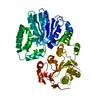 4v38C  4v39C 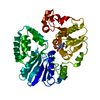 4v3bC 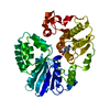 4v3cC  2ilvS C: citing same article ( S: Starting model for refinement |
|---|---|
| Similar structure data |


-Links
 PDBj
PDBj- Assembly
| Deposited unit | 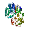
| ||||||||
|---|---|---|---|---|---|---|---|---|---|
| 1 |
| ||||||||
| Unit cell |
|
-Components
| #1: Protein | Mass: 45662.023 Da / Num. of mol.: 1 Source method: isolated from a genetically manipulated source Source: (gene. exp.) PASTEURELLA DAGMATIS (bacteria) / Production host: ESCHERICHIA COLI (E. coli) / References: UniProt: K9UUI6 |
|---|---|
| #2: Water | ChemComp-HOH / Water Mass: 18.015 Da / Num. of mol.: 11 / Source method: isolated from a natural source / Formula: H2O Mass: 18.015 Da / Num. of mol.: 11 / Source method: isolated from a natural source / Formula: H2O |
-Experimental details
-Experiment
| Experiment | Method: X-RAY DIFFRACTION / Number of used crystals: 1 |
|---|
- Sample preparation
| Crystal | Density Matthews: 2.6 Å3/Da / Density % sol: 53 % / Description: NONE |
|---|---|
| Crystal grow | Details: 3.3 MG/ML OF PROTEIN INDEX SCREEN CONDITION D9 (0.1M TRIS PH 8.5, 25% W/V POLYETHYLENE GLYCOL 3350) |
-Data collection
| Diffraction | Mean temperature: 100 K |
|---|---|
| Diffraction source | Source: SYNCHROTRON / Site: PETRA III, DESY  / Beamline: P11 / Wavelength: 1.0332 / Beamline: P11 / Wavelength: 1.0332 |
| Detector | Type: PILATUS / Detector: PIXEL / Date: Jan 29, 2014 |
| Radiation | Protocol: SINGLE WAVELENGTH / Monochromatic (M) / Laue (L): M / Scattering type: x-ray |
| Radiation wavelength | Wavelength: 1.0332 Å / Relative weight: 1 |
| Reflection | Resolution: 2.71→45.1 Å / Num. obs: 12618 / % possible obs: 98.8 % / Observed criterion σ(I): 1.9 / Redundancy: 3.3 % / Rmerge(I) obs: 0.11 / Net I/σ(I): 10.4 |
| Reflection shell | Resolution: 2.71→2.87 Å / Redundancy: 3.2 % / Rmerge(I) obs: 0.63 / Mean I/σ(I) obs: 1.9 / % possible all: 97.3 |
- Processing
| Software |
| ||||||||||||||||||||||||||||||||||||||||||||||||||||||||||||||||||||||||||||||||||||||||||||||||||||||||||||||||||||||||||||||||||||||||||||||||||||||||||||||||||||||||||||||||||||||
|---|---|---|---|---|---|---|---|---|---|---|---|---|---|---|---|---|---|---|---|---|---|---|---|---|---|---|---|---|---|---|---|---|---|---|---|---|---|---|---|---|---|---|---|---|---|---|---|---|---|---|---|---|---|---|---|---|---|---|---|---|---|---|---|---|---|---|---|---|---|---|---|---|---|---|---|---|---|---|---|---|---|---|---|---|---|---|---|---|---|---|---|---|---|---|---|---|---|---|---|---|---|---|---|---|---|---|---|---|---|---|---|---|---|---|---|---|---|---|---|---|---|---|---|---|---|---|---|---|---|---|---|---|---|---|---|---|---|---|---|---|---|---|---|---|---|---|---|---|---|---|---|---|---|---|---|---|---|---|---|---|---|---|---|---|---|---|---|---|---|---|---|---|---|---|---|---|---|---|---|---|---|---|---|
| Refinement | Method to determine structure: MOLECULAR REPLACEMENT Starting model: PDB ENTRY 2ILV Resolution: 2.71→45.11 Å / Cor.coef. Fo:Fc: 0.95 / Cor.coef. Fo:Fc free: 0.898 / SU B: 16.81 / SU ML: 0.324 / Cross valid method: THROUGHOUT / ESU R Free: 0.374 / Stereochemistry target values: MAXIMUM LIKELIHOOD Details: HYDROGENS HAVE BEEN ADDED IN THE RIDING POSITIONS. U VALUES, REFINED INDIVIDUALLY RESIDUES 36-39 ARE DISORDERED OR SHOW MULTIPLE CONFORMATIONS AND COULD NOT BE BUILD WITH CERTAINTY
| ||||||||||||||||||||||||||||||||||||||||||||||||||||||||||||||||||||||||||||||||||||||||||||||||||||||||||||||||||||||||||||||||||||||||||||||||||||||||||||||||||||||||||||||||||||||
| Solvent computation | Ion probe radii: 0.8 Å / Shrinkage radii: 0.8 Å / VDW probe radii: 1.2 Å / Solvent model: MASK | ||||||||||||||||||||||||||||||||||||||||||||||||||||||||||||||||||||||||||||||||||||||||||||||||||||||||||||||||||||||||||||||||||||||||||||||||||||||||||||||||||||||||||||||||||||||
| Displacement parameters | Biso mean: 46.174 Å2
| ||||||||||||||||||||||||||||||||||||||||||||||||||||||||||||||||||||||||||||||||||||||||||||||||||||||||||||||||||||||||||||||||||||||||||||||||||||||||||||||||||||||||||||||||||||||
| Refinement step | Cycle: LAST / Resolution: 2.71→45.11 Å
| ||||||||||||||||||||||||||||||||||||||||||||||||||||||||||||||||||||||||||||||||||||||||||||||||||||||||||||||||||||||||||||||||||||||||||||||||||||||||||||||||||||||||||||||||||||||
| Refine LS restraints |
|