- PDB-7s7j: Structure of Human SPASTIN-IST1 complex. -
+
Open data
ID or keywords:
Loading...
-
Basic information
Entry
Database: PDB / ID: 7s7j
Title
Structure of Human SPASTIN-IST1 complex.
Components
IST1 homolog
Spastin
Keywords
PROTEIN TRANSPORT / cytokinesis / MIT / AAA-ATPase
Function / homology
Function and homology information
cytokinetic process / microtubule-severing ATPase / microtubule severing ATPase activity / viral capsid secondary envelopment / microtubule severing / MIT domain binding / endoplasmic reticulum tubular network / positive regulation of microtubule depolymerization / abscission / ESCRT III complex disassembly ...cytokinetic process / microtubule-severing ATPase / microtubule severing ATPase activity / viral capsid secondary envelopment / microtubule severing / MIT domain binding / endoplasmic reticulum tubular network / positive regulation of microtubule depolymerization / abscission / ESCRT III complex disassembly / cytoskeleton-dependent cytokinesis / mitotic nuclear membrane reassembly / collateral sprouting / nuclear membrane reassembly / Sealing of the nuclear envelope (NE) by ESCRT-III / positive regulation of collateral sprouting / membrane fission / exit from mitosis / multivesicular body assembly / microtubule bundle formation / anterograde axonal transport / mitotic spindle disassembly / protein hexamerization / Flemming body / axonal transport of mitochondrion / beta-tubulin binding / positive regulation of cytokinesis / positive regulation of proteolysis / mitotic cytokinesis / alpha-tubulin binding / viral release from host cell / endoplasmic reticulum-Golgi intermediate compartment / metabolic process / endoplasmic reticulum to Golgi vesicle-mediated transport / axon cytoplasm / isomerase activity / lipid droplet / axonogenesis / establishment of protein localization / protein homooligomerization / protein localization / spindle pole / azurophil granule lumen / protein transport / nuclear envelope / midbody / cytoplasmic vesicle / microtubule binding / nuclear membrane / microtubule / endosome / cadherin binding / protein domain specific binding / axon / cell division / intracellular membrane-bounded organelle / centrosome / chromatin / Neutrophil degranulation / protein-containing complex binding / endoplasmic reticulum membrane / perinuclear region of cytoplasm / ATP hydrolysis activity / extracellular exosome / extracellular region / nucleoplasm / ATP binding / identical protein binding / nucleus / cytosol / cytoplasm Similarity search - Function
Spastin, chordate / Spastin / Vacuolar protein sorting-associated protein Ist1 / Regulator of Vps4 activity in the MVB pathway / Vacuolar protein sorting-associated protein IST1-like / Vps4 oligomerisation, C-terminal / MIT domain / Microtubule Interacting and Trafficking molecule domain / Vps4 C terminal oligomerisation domain / AAA ATPase, AAA+ lid domain ...Spastin, chordate / Spastin / Vacuolar protein sorting-associated protein Ist1 / Regulator of Vps4 activity in the MVB pathway / Vacuolar protein sorting-associated protein IST1-like / Vps4 oligomerisation, C-terminal / MIT domain / Microtubule Interacting and Trafficking molecule domain / Vps4 C terminal oligomerisation domain / AAA ATPase, AAA+ lid domain / AAA+ lid domain / ATPase, AAA-type, conserved site / AAA-protein family signature. / ATPase family associated with various cellular activities (AAA) / ATPase, AAA-type, core / ATPases associated with a variety of cellular activities / AAA+ ATPase domain / P-loop containing nucleoside triphosphate hydrolase Similarity search - Domain/homology
In the structure databanks used in Yorodumi, some data are registered as the other names, "COVID-19 virus" and "2019-nCoV". Here are the details of the virus and the list of structure data.
Jan 31, 2019. EMDB accession codes are about to change! (news from PDBe EMDB page)
EMDB accession codes are about to change! (news from PDBe EMDB page)
The allocation of 4 digits for EMDB accession codes will soon come to an end. Whilst these codes will remain in use, new EMDB accession codes will include an additional digit and will expand incrementally as the available range of codes is exhausted. The current 4-digit format prefixed with “EMD-” (i.e. EMD-XXXX) will advance to a 5-digit format (i.e. EMD-XXXXX), and so on. It is currently estimated that the 4-digit codes will be depleted around Spring 2019, at which point the 5-digit format will come into force.
The EM Navigator/Yorodumi systems omit the EMD- prefix.
Related info.:Q: What is EMD? / ID/Accession-code notation in Yorodumi/EM Navigator
Yorodumi is a browser for structure data from EMDB, PDB, SASBDB, etc.
This page is also the successor to EM Navigator detail page, and also detail information page/front-end page for Omokage search.
The word "yorodu" (or yorozu) is an old Japanese word meaning "ten thousand". "mi" (miru) is to see.
Related info.:EMDB / PDB / SASBDB / Comparison of 3 databanks / Yorodumi Search / Aug 31, 2016. New EM Navigator & Yorodumi / Yorodumi Papers / Jmol/JSmol / Function and homology information / Changes in new EM Navigator and Yorodumi
 Movie
Movie Controller


 Open data
 Basic information
 Components
 Keywords
 PROTEIN TRANSPORT /
  Function and homology information
 Authors
 United States, 1items
  Citation
 Structure visualization
 Downloads & links
 Other downloads
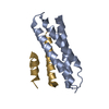
 PDBj
PDBj


 Assembly


 Mass: 194.226 Da / Num. of mol.: 1 / Source method: obtained synthetically / Formula: C8H18O5 / Comment: precipitant*YM
 Mass: 40.078 Da / Num. of mol.: 1 / Source method: obtained synthetically / Formula: Ca
 Mass: 150.173 Da / Num. of mol.: 1 / Source method: obtained synthetically / Formula: C6H14O4
 Mass: 35.453 Da / Num. of mol.: 1 / Source method: obtained synthetically / Formula: Cl
 Sample preparation
 Processing