 Movie
Movie Controller

[English] 日本語
 Yorodumi
- PDB-7s68: Structure of human PARP1 domains (Zn1, Zn3, WGR and HD) bound to ... -
+ Open data

- Basic information
| Entry | Database: PDB / ID: 7s68 | ||||||
|---|---|---|---|---|---|---|---|
| Title | Structure of human PARP1 domains (Zn1, Zn3, WGR and HD) bound to a DNA double strand break. | ||||||
 Components Components |
| ||||||
 Keywords Keywords | DNA BINDING PROTEIN/DNA / PARP / ADP-ribose transferase / DNA break detection /  zinc finger / DNA BINDING PROTEIN / DNA BINDING PROTEIN-DNA complex zinc finger / DNA BINDING PROTEIN / DNA BINDING PROTEIN-DNA complex | ||||||
| Function / homology |  Function and homology information Function and homology informationNAD+-histone H2BS6 serine ADP-ribosyltransferase activity / NAD+-histone H3S10 serine ADP-ribosyltransferase activity / NAD+-histone H2BE35 glutamate ADP-ribosyltransferase activity / regulation of base-excision repair / positive regulation of myofibroblast differentiation / negative regulation of ATP biosynthetic process / NAD+-protein-tyrosine ADP-ribosyltransferase activity / NAD+-protein-histidine ADP-ribosyltransferase activity / carbohydrate biosynthetic process / regulation of circadian sleep/wake cycle, non-REM sleep ...NAD+-histone H2BS6 serine ADP-ribosyltransferase activity / NAD+-histone H3S10 serine ADP-ribosyltransferase activity / NAD+-histone H2BE35 glutamate ADP-ribosyltransferase activity / regulation of base-excision repair / positive regulation of myofibroblast differentiation / negative regulation of ATP biosynthetic process / NAD+-protein-tyrosine ADP-ribosyltransferase activity / NAD+-protein-histidine ADP-ribosyltransferase activity / carbohydrate biosynthetic process / regulation of circadian sleep/wake cycle, non-REM sleep / positive regulation of single strand break repair / vRNA Synthesis / negative regulation of adipose tissue development / NAD+-protein-serine ADP-ribosyltransferase activity / NAD DNA ADP-ribosyltransferase activity / NAD+- protein-aspartate ADP-ribosyltransferase activity / NAD+-protein-glutamate ADP-ribosyltransferase activity / mitochondrial DNA metabolic process / DNA ADP-ribosylation / regulation of oxidative stress-induced neuron intrinsic apoptotic signaling pathway / signal transduction involved in regulation of gene expression / positive regulation of necroptotic process / regulation of catalytic activity / ATP generation from poly-ADP-D-ribose / replication fork reversal / transcription regulator activator activity / HDR through MMEJ (alt-NHEJ) / positive regulation of DNA-templated transcription, elongation / NAD+ ADP-ribosyltransferase / cellular response to zinc ion / negative regulation of telomere maintenance via telomere lengthening / positive regulation of intracellular estrogen receptor signaling pathway / protein auto-ADP-ribosylation / positive regulation of mitochondrial depolarization / response to aldosterone / mitochondrial DNA repair / negative regulation of cGAS/STING signaling pathway / protein poly-ADP-ribosylation / positive regulation of cardiac muscle hypertrophy / negative regulation of transcription elongation by RNA polymerase II / nuclear replication fork / site of DNA damage / NAD+-protein ADP-ribosyltransferase activity / R-SMAD binding / positive regulation of SMAD protein signal transduction / protein autoprocessing / macrophage differentiation / decidualization / Transferases; Glycosyltransferases; Pentosyltransferases / positive regulation of double-strand break repair via homologous recombination / NAD+ ADP-ribosyltransferase activity / POLB-Dependent Long Patch Base Excision Repair / nucleosome binding / SUMOylation of DNA damage response and repair proteins / protein localization to chromatin / negative regulation of innate immune response / telomere maintenance / nucleotidyltransferase activity / mitochondrion organization / transforming growth factor beta receptor signaling pathway / cellular response to nerve growth factor stimulus / nuclear estrogen receptor binding / protein-DNA complex / response to gamma radiation / Downregulation of SMAD2/3:SMAD4 transcriptional activity / DNA Damage Recognition in GG-NER / protein modification process / Dual Incision in GG-NER / Formation of Incision Complex in GG-NER / histone deacetylase binding / positive regulation of protein localization to nucleus / cellular response to insulin stimulus / cellular response to amyloid-beta / NAD binding / cellular response to UV / regulation of protein localization / double-strand break repair / nuclear envelope / site of double-strand break / cellular response to oxidative stress / positive regulation of canonical NF-kappaB signal transduction / RNA polymerase II-specific DNA-binding transcription factor binding / transcription regulator complex / transcription by RNA polymerase II / damaged DNA binding / chromosome, telomeric region / nuclear body / DNA repair / innate immune response / negative regulation of DNA-templated transcription / apoptotic process / DNA damage response / ubiquitin protein ligase binding / chromatin binding / chromatin / nucleolus / protein kinase binding / enzyme binding / negative regulation of transcription by RNA polymerase II / protein homodimerization activitySimilarity search - Function | ||||||
| Biological species |  Homo sapiens (human)DNA molecule (others) Homo sapiens (human)DNA molecule (others) | ||||||
| Method | X-RAY DIFFRACTION / SYNCHROTRON / MOLECULAR REPLACEMENT / Resolution: 3.3 Å | ||||||
 Authors Authors | Rouleau-Turcotte, E. / Pascal, J.M. | ||||||
| Funding support |  Canada, 1items Canada, 1items
| ||||||
 Citation Citation | Journal: Mol.Cell / Year: 2022 Title: Captured snapshots of PARP1 in the active state reveal the mechanics of PARP1 allostery. Authors: Rouleau-Turcotte, E. / Krastev, D.B. / Pettitt, S.J. / Lord, C.J. / Pascal, J.M. | ||||||
| History |
|
- Structure visualization
| Structure viewer | Molecule: MolmilJmol/JSmol |
|---|
- Downloads & links
-Download
| PDBx/mmCIF format | 7s68.cif.gz | 300.4 KB | Display | PDBx/mmCIF format |
|---|---|---|---|---|
| PDB format | pdb7s68.ent.gz | 243.1 KB | Display | PDB format |
| PDBx/mmJSON format | 7s68.json.gz | Tree view | PDBx/mmJSON format | |
| Others |  Other downloads Other downloads |
-Validation report
| Arichive directory | https://data.pdbj.org/pub/pdb/validation_reports/s6/7s68ftp://data.pdbj.org/pub/pdb/validation_reports/s6/7s68 | HTTPS FTP |
|---|
-Related structure data
| Related structure data |  7s6hC 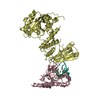 7s6mC  7s81C  4dqyS C: citing same article ( S: Starting model for refinement |
|---|---|
| Similar structure data |
-Links
 PDBj
PDBj


- Assembly
| Deposited unit | 
| ||||||||
|---|---|---|---|---|---|---|---|---|---|
| 1 |
| ||||||||
| Unit cell |
|
-Components
| #1: DNA chain | Mass: 3045.992 Da / Num. of mol.: 2 / Source method: obtained synthetically / Source: (synth.) DNA molecule (others)#2: Protein | | Mass: 29813.668 Da / Num. of mol.: 1 Source method: isolated from a genetically manipulated source Source: (gene. exp.) Homo sapiens (human) / Gene: PARP1, ADPRT, PPOL / Production host:  Escherichia coli (E. coli) Escherichia coli (E. coli)References: UniProt: P09874, NAD+ ADP-ribosyltransferase, Transferases; Glycosyltransferases; Pentosyltransferases#3: Protein | | Mass: 31262.848 Da / Num. of mol.: 1 Source method: isolated from a genetically manipulated source Source: (gene. exp.) Homo sapiens (human) / Gene: PARP1, ADPRT, PPOL / Production host: Escherichia coli (E. coli)References: UniProt: P09874, NAD+ ADP-ribosyltransferase, Transferases; Glycosyltransferases; Pentosyltransferases#4: Chemical |   Mass: 65.409 Da / Num. of mol.: 2 / Source method: obtained synthetically / Formula: Zn Mass: 65.409 Da / Num. of mol.: 2 / Source method: obtained synthetically / Formula: ZnHas ligand of interest | N | |
|---|
-Experimental details
-Experiment
| Experiment | Method: X-RAY DIFFRACTION / Number of used crystals: 1 |
|---|
- Sample preparation
| Crystal | Density Matthews: 3.48 Å3/Da / Density % sol: 64.65 % |
|---|---|
| Crystal grow | Temperature: 293 K / Method: vapor diffusion, sitting drop / Details: PEG 8000, ethylene glycol, HEPES pH 7.5 |
-Data collection
| Diffraction | Mean temperature: 100 K / Serial crystal experiment: N | ||||||||||||||||||||||||||||||
|---|---|---|---|---|---|---|---|---|---|---|---|---|---|---|---|---|---|---|---|---|---|---|---|---|---|---|---|---|---|---|---|
| Diffraction source | Source: SYNCHROTRON / Site: ALS  / Beamline: 8.3.1 / Wavelength: 0.98 Å / Beamline: 8.3.1 / Wavelength: 0.98 Å | ||||||||||||||||||||||||||||||
| Detector | Type: DECTRIS PILATUS3 S 6M / Detector: PIXEL / Date: Oct 17, 2019 | ||||||||||||||||||||||||||||||
| Radiation | Protocol: SINGLE WAVELENGTH / Monochromatic (M) / Laue (L): M / Scattering type: x-ray | ||||||||||||||||||||||||||||||
| Radiation wavelength | Wavelength: 0.98 Å / Relative weight: 1 | ||||||||||||||||||||||||||||||
| Reflection | Resolution: 3.3→47.41 Å / Num. obs: 13906 / % possible obs: 99.3 % / Redundancy: 8.3 % / CC1/2: 0.999 / Rmerge(I) obs: 0.096 / Rpim(I) all: 0.036 / Rrim(I) all: 0.103 / Net I/σ(I): 14.1 / Num. measured all: 115298 / Scaling rejects: 151 | ||||||||||||||||||||||||||||||
| Reflection shell | Diffraction-ID: 1
|
- Processing
| Software |
| ||||||||||||||||||||||||||||||||||||||||||||||||||||||||||||||||||||||||||||||||||||||||||||||||||||||||||||||||||||||||||||||||||||||||||||||||||||||
|---|---|---|---|---|---|---|---|---|---|---|---|---|---|---|---|---|---|---|---|---|---|---|---|---|---|---|---|---|---|---|---|---|---|---|---|---|---|---|---|---|---|---|---|---|---|---|---|---|---|---|---|---|---|---|---|---|---|---|---|---|---|---|---|---|---|---|---|---|---|---|---|---|---|---|---|---|---|---|---|---|---|---|---|---|---|---|---|---|---|---|---|---|---|---|---|---|---|---|---|---|---|---|---|---|---|---|---|---|---|---|---|---|---|---|---|---|---|---|---|---|---|---|---|---|---|---|---|---|---|---|---|---|---|---|---|---|---|---|---|---|---|---|---|---|---|---|---|---|---|---|---|
| Refinement | Method to determine structure: MOLECULAR REPLACEMENT Starting model: 4DQY Resolution: 3.3→47.41 Å / SU ML: 0.94 / Cross valid method: THROUGHOUT / σ(F): 1.34 / Phase error: 48.48 / Stereochemistry target values: ML
| ||||||||||||||||||||||||||||||||||||||||||||||||||||||||||||||||||||||||||||||||||||||||||||||||||||||||||||||||||||||||||||||||||||||||||||||||||||||
| Solvent computation | Shrinkage radii: 0.9 Å / VDW probe radii: 1.11 Å / Solvent model: FLAT BULK SOLVENT MODEL | ||||||||||||||||||||||||||||||||||||||||||||||||||||||||||||||||||||||||||||||||||||||||||||||||||||||||||||||||||||||||||||||||||||||||||||||||||||||
| Displacement parameters | Biso max: 388.52 Å2 / Biso mean: 169.7006 Å2 / Biso min: 64.83 Å2 | ||||||||||||||||||||||||||||||||||||||||||||||||||||||||||||||||||||||||||||||||||||||||||||||||||||||||||||||||||||||||||||||||||||||||||||||||||||||
| Refinement step | Cycle: final / Resolution: 3.3→47.41 Å
| ||||||||||||||||||||||||||||||||||||||||||||||||||||||||||||||||||||||||||||||||||||||||||||||||||||||||||||||||||||||||||||||||||||||||||||||||||||||
| LS refinement shell | Refine-ID: X-RAY DIFFRACTION / Rfactor Rfree error: 0 / Total num. of bins used: 4
| ||||||||||||||||||||||||||||||||||||||||||||||||||||||||||||||||||||||||||||||||||||||||||||||||||||||||||||||||||||||||||||||||||||||||||||||||||||||
| Refinement TLS params. | Method: refined / Refine-ID: X-RAY DIFFRACTION
| ||||||||||||||||||||||||||||||||||||||||||||||||||||||||||||||||||||||||||||||||||||||||||||||||||||||||||||||||||||||||||||||||||||||||||||||||||||||
| Refinement TLS group |
|
