 Movie
Movie Controller

[English] 日本語
 Yorodumi
- PDB-7s4m: CryoEM structure of Methylocystis sp. str. Rockwell pMMO in a POP... -
+ Open data

- Basic information
| Entry | Database: PDB / ID: 7s4m | ||||||
|---|---|---|---|---|---|---|---|
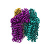
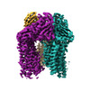
| Title | CryoEM structure of Methylocystis sp. str. Rockwell pMMO in a POPC nanodisc at 2.42 Angstrom resolution | ||||||
 Components Components |
| ||||||
 Keywords Keywords |  OXIDOREDUCTASE / Complex OXIDOREDUCTASE / Complex | ||||||
| Function / homology | COPPER (II) ION / DECANE / DODECANE / 1,2-dihexanoyl-sn-glycero-3-phosphocholine Function and homology information Function and homology information | ||||||
| Biological species |  Methylocystis sp. ATCC 49242 (bacteria) Methylocystis sp. ATCC 49242 (bacteria) | ||||||
| Method | ELECTRON MICROSCOPY / single particle reconstruction / cryo EM / Resolution: 2.42 Å | ||||||
 Authors Authors | Koo, C.W. / Rosenzweig, A.C. | ||||||
| Funding support |  United States, 1items United States, 1items
| ||||||
 Citation Citation | Journal: Science / Year: 2022 Title: Recovery of particulate methane monooxygenase structure and activity in a lipid bilayer. Authors: Christopher W Koo / Frank J Tucci / Yuan He / Amy C Rosenzweig / Abstract: Bacterial methane oxidation using the enzyme particulate methane monooxygenase (pMMO) contributes to the removal of environmental methane, a potent greenhouse gas. Crystal structures determined using ...Bacterial methane oxidation using the enzyme particulate methane monooxygenase (pMMO) contributes to the removal of environmental methane, a potent greenhouse gas. Crystal structures determined using inactive, detergent-solubilized pMMO lack several conserved regions neighboring the proposed active site. We show that reconstituting pMMO in nanodiscs with lipids extracted from the native organism restores methane oxidation activity. Multiple nanodisc-embedded pMMO structures determined by cryo-electron microscopy to 2.14- to 2.46-angstrom resolution reveal the structure of pMMO in a lipid environment. The resulting model includes stabilizing lipids, regions of the PmoA and PmoC subunits not observed in prior structures, and a previously undetected copper-binding site in the PmoC subunit with an adjacent hydrophobic cavity. These structures provide a revised framework for understanding and engineering pMMO function. | ||||||
| History |
|
- Structure visualization
| Structure viewer | Molecule: MolmilJmol/JSmol |
|---|
- Downloads & links
-Download
| PDBx/mmCIF format | 7s4m.cif.gz | 488.1 KB | Display | PDBx/mmCIF format |
|---|---|---|---|---|
| PDB format | pdb7s4m.ent.gz | 406.7 KB | Display | PDB format |
| PDBx/mmJSON format | 7s4m.json.gz | Tree view | PDBx/mmJSON format | |
| Others |  Other downloads Other downloads |
-Validation report
| Arichive directory | https://data.pdbj.org/pub/pdb/validation_reports/s4/7s4mftp://data.pdbj.org/pub/pdb/validation_reports/s4/7s4m | HTTPS FTP |
|---|
-Related structure data
| Related structure data |  24831MC 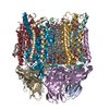 7s4hC  7s4iC 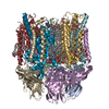 7s4jC  7s4kC  7s4lC 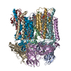 7t4oC  7t4pC M: map data used to model this data C: citing same article ( |
|---|---|
| Similar structure data |


-Links
 PDBj
PDBj- Assembly
| Deposited unit | 
|
|---|---|
| 1 |
|
-Components
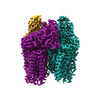
-Particulate methane monooxygenase ... , 2 types, 6 molecules AEIFBJ
| #1: Protein | Mass: 42785.566 Da / Num. of mol.: 3 / Source method: isolated from a natural source / Source: (natural) Methylocystis sp. ATCC 49242 (bacteria)#2: Protein | Mass: 27796.467 Da / Num. of mol.: 3 / Source method: isolated from a natural source / Source: (natural) Methylocystis sp. ATCC 49242 (bacteria) |
|---|
-Protein / Protein/peptide , 2 types, 6 molecules GCKDNH
| #3: Protein | Mass: 27924.629 Da / Num. of mol.: 3 / Source method: isolated from a natural source / Source: (natural) Methylocystis sp. ATCC 49242 (bacteria)#4: Protein/peptide | Mass: 1635.006 Da / Num. of mol.: 3 / Source method: isolated from a natural source / Source: (natural) Methylocystis sp. ATCC 49242 (bacteria) |
|---|
-Non-polymers , 5 types, 63 molecules 


| #5: Chemical | ChemComp-CU / Copper Mass: 63.546 Da / Num. of mol.: 6 / Source method: obtained synthetically / Formula: Cu / Feature type: SUBJECT OF INVESTIGATION Mass: 63.546 Da / Num. of mol.: 6 / Source method: obtained synthetically / Formula: Cu / Feature type: SUBJECT OF INVESTIGATION#6: Chemical | ChemComp-HXG /  Mass: 454.515 Da / Num. of mol.: 12 / Source method: isolated from a natural source / Formula: C20H41NO8P / Feature type: SUBJECT OF INVESTIGATION Mass: 454.515 Da / Num. of mol.: 12 / Source method: isolated from a natural source / Formula: C20H41NO8P / Feature type: SUBJECT OF INVESTIGATION#7: Chemical | ChemComp-D10 / Decane Mass: 142.282 Da / Num. of mol.: 6 / Source method: obtained synthetically / Formula: C10H22 / Feature type: SUBJECT OF INVESTIGATION Mass: 142.282 Da / Num. of mol.: 6 / Source method: obtained synthetically / Formula: C10H22 / Feature type: SUBJECT OF INVESTIGATION#8: Chemical | Dodecane Mass: 170.335 Da / Num. of mol.: 3 / Source method: obtained synthetically / Formula: C12H26 / Feature type: SUBJECT OF INVESTIGATION Mass: 170.335 Da / Num. of mol.: 3 / Source method: obtained synthetically / Formula: C12H26 / Feature type: SUBJECT OF INVESTIGATION#9: Water | ChemComp-HOH / | WaterMass: 18.015 Da / Num. of mol.: 36 / Source method: isolated from a natural source / Formula: H2O |
|---|
-Details
| Has ligand of interest | Y |
|---|
-Experimental details
-Experiment
| Experiment | Method: ELECTRON MICROSCOPY |
|---|---|
| EM experiment | Aggregation state: PARTICLE / 3D reconstruction method: single particle reconstruction |
- Sample preparation
| Component | Name: pMMO complex in a native lipid nanodisc / Type: COMPLEX / Entity ID: #1-#4 / Source: NATURAL |
|---|---|
| Source (natural) | Organism: Methylocystis sp. ATCC 49242 (bacteria) |
| Buffer solution | pH: 7.3 |
| Specimen | Embedding applied: NO / Shadowing applied: NO / Staining applied: NO / Vitrification applied: YES |
| Vitrification | Cryogen name: ETHANE |
- Electron microscopy imaging
| Experimental equipment |  Model: Titan Krios / Image courtesy: FEI Company |
|---|---|
| Microscopy | Model: FEI TITAN KRIOS |
| Electron gun | Electron source: FIELD EMISSION GUN / Accelerating voltage: 300 kV / Illumination mode: FLOOD BEAM |
| Electron lens | Mode: BRIGHT FIELDBright-field microscopy |
| Image recording | Electron dose: 1.02 e/Å2 / Film or detector model: GATAN K3 (6k x 4k) |
- Processing
| CTF correction | Type: PHASE FLIPPING AND AMPLITUDE CORRECTION |
|---|---|
| 3D reconstruction | Resolution: 2.42 Å / Resolution method: FSC 0.143 CUT-OFF / Num. of particles: 490196 / Symmetry type: POINT |
